Copyright
©The Author(s) 2021.
World J Clin Cases. Sep 16, 2021; 9(26): 7825-7832
Published online Sep 16, 2021. doi: 10.12998/wjcc.v9.i26.7825
Published online Sep 16, 2021. doi: 10.12998/wjcc.v9.i26.7825
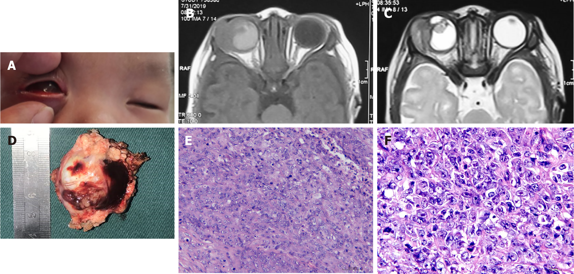
Figure 1 A 1-mo-old male newborn.
A: A mass located in the right eye, with proptosis; B and C: Axial T-1 weighted (B) and T-2 weighted (C) magnetic resonance images of the orbit revealed a large mass near the lateral orbital wall with slightly shorter T1 and shorter T2 signals. The eyeball structure is unclear, with abnormal signals; D: Gross morphology of the tumor showing the tumor tissue around the eyeball; E and F: Histopathologic analysis including micrographs after hematoxylin-eosin (E, × 20; F, × 40). A small, round, dark blue tumor with a monotonous, highly cellular pattern was observed with tumor cell atypia, and poorly differentiated cells.
- Citation: Zhang Y, Li YY, Yu HY, Xie XL, Zhang HM, He F, Li HY. Rare neonatal malignant primary orbital tumors: Three case reports. World J Clin Cases 2021; 9(26): 7825-7832
- URL: https://www.wjgnet.com/2307-8960/full/v9/i26/7825.htm
- DOI: https://dx.doi.org/10.12998/wjcc.v9.i26.7825


