Copyright
©The Author(s) 2021.
World J Clin Cases. Sep 6, 2021; 9(25): 7588-7592
Published online Sep 6, 2021. doi: 10.12998/wjcc.v9.i25.7588
Published online Sep 6, 2021. doi: 10.12998/wjcc.v9.i25.7588
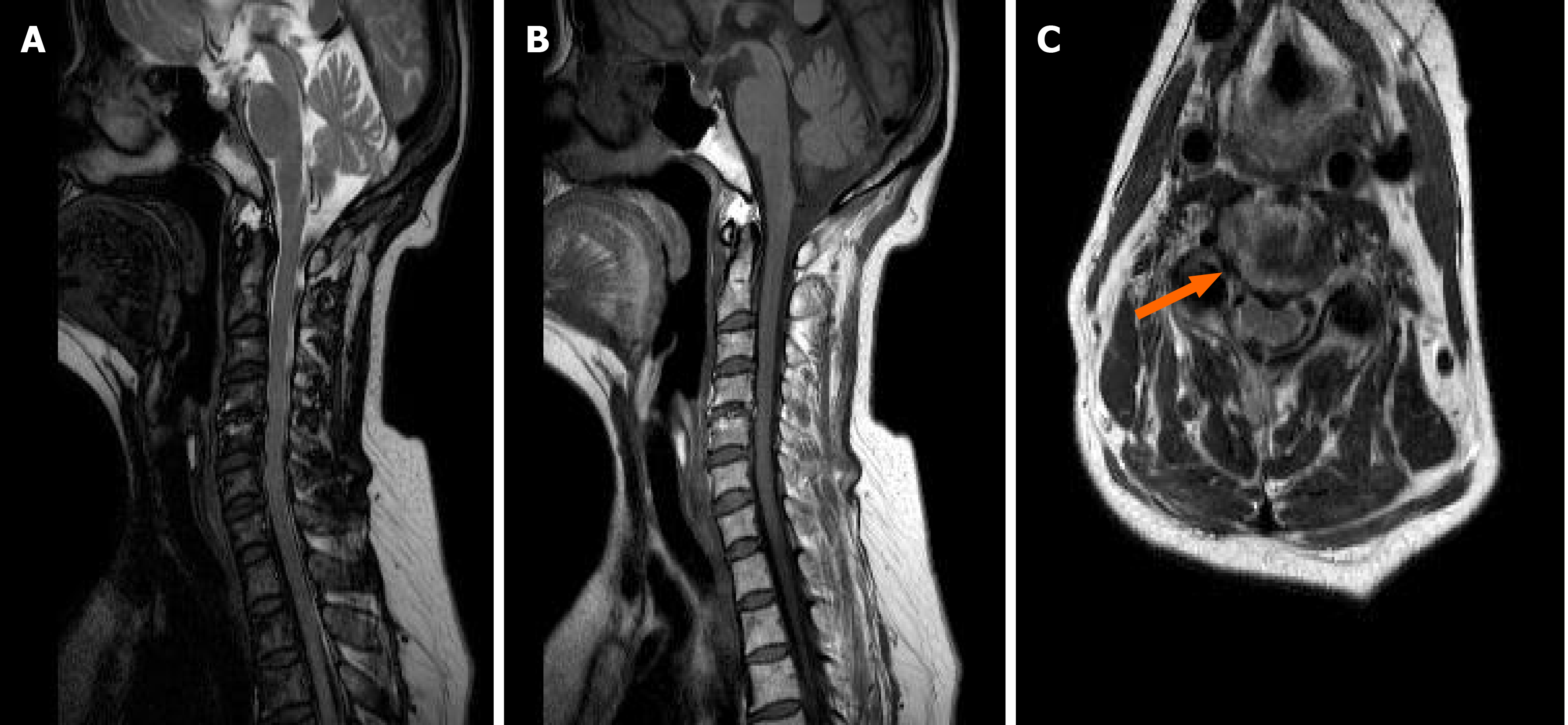
Figure 2 Cervical spine magnetic resonance images.
A: T2-weighted magnetic resonance (MR) image of the cervical spine (sagittal view) showing intervertebral disc protrusion of C3-4-5 segments; B: T1-weighted MR image of the cervical spine (sagittal view) showing intervertebral disc protrusion of C3-4-5; C: T2-weighted MR image of the cervical spine (transactional view) showing degeneration of the right facet joint of C4-5 segment (orange arrow).
- Citation: Yun G, Kim E, Baik J, Do W, Jung YH, You CM. Diagnosis and management of ophthalmic zoster sine herpete accompanied by cervical spine disc protrusion: A case report. World J Clin Cases 2021; 9(25): 7588-7592
- URL: https://www.wjgnet.com/2307-8960/full/v9/i25/7588.htm
- DOI: https://dx.doi.org/10.12998/wjcc.v9.i25.7588


