Copyright
©The Author(s) 2021.
World J Clin Cases. Sep 6, 2021; 9(25): 7579-7587
Published online Sep 6, 2021. doi: 10.12998/wjcc.v9.i25.7579
Published online Sep 6, 2021. doi: 10.12998/wjcc.v9.i25.7579
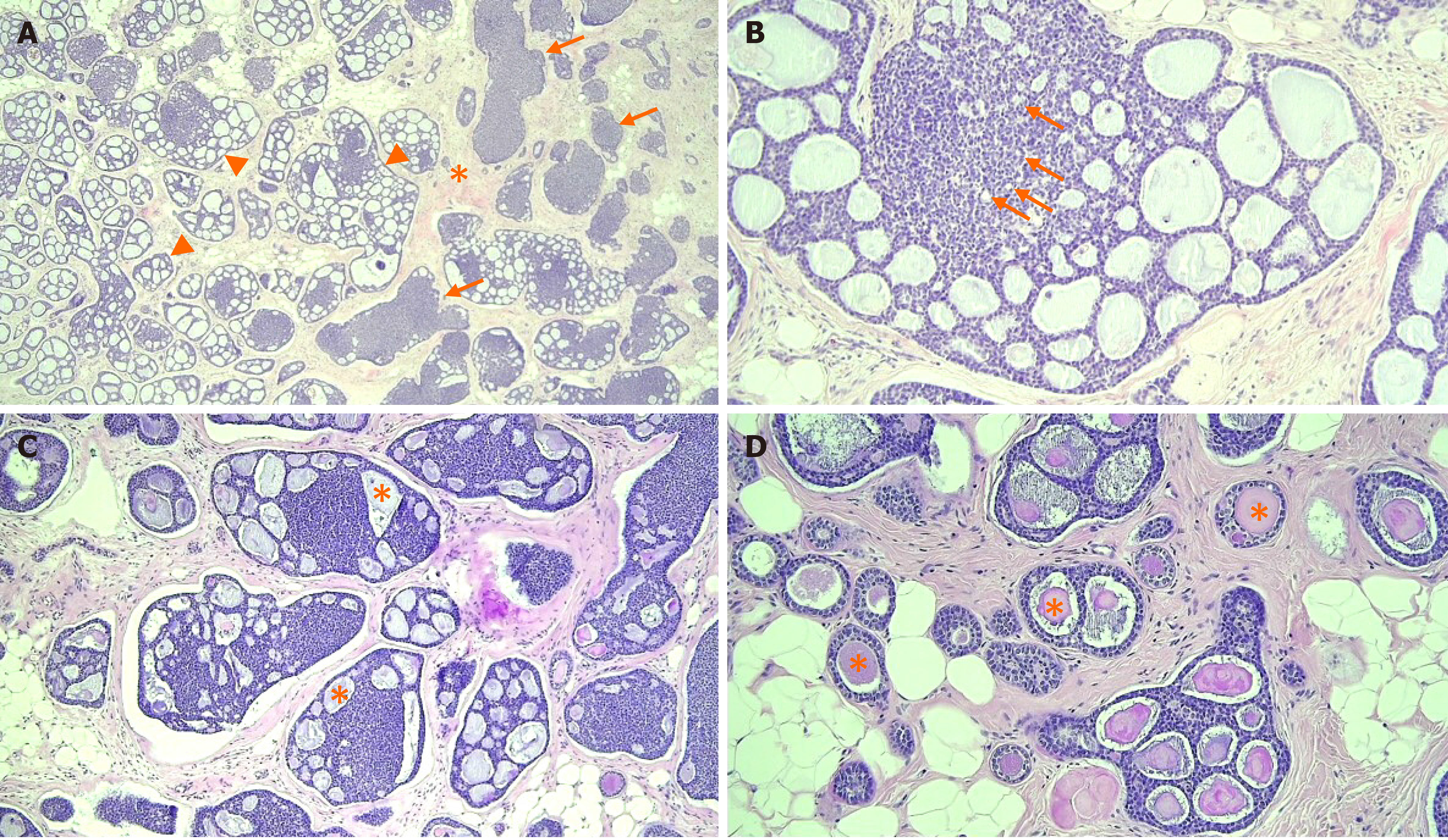
Figure 4 Photomicrographs of the adenoid cystic carcinoma.
A: The tumor was composed of cribriform (arrowheads) and solid nests (arrows) in the fibrous stroma (asterisk), [hematoxylin and eosin (H&E) staining, magnification: 40 ×]; B: Myoepithelial cells formed cribriform or solid basaloid growth, whereas ductal epithelial cells were lining small ductule-like lumen (arrows) (H&E staining, magnification: 200 ×); C and D: The cribriform space contained Periodic acid Schiff positive basement membrane-like materials (asterisk) in invasive (H&E staining, magnification: 100 ×) and in situ components (H&E staining, magnification: 200 ×).
- Citation: An JK, Woo JJ, Kim EK, Kwak HY. Breast adenoid cystic carcinoma arising in microglandular adenosis: A case report and review of literature. World J Clin Cases 2021; 9(25): 7579-7587
- URL: https://www.wjgnet.com/2307-8960/full/v9/i25/7579.htm
- DOI: https://dx.doi.org/10.12998/wjcc.v9.i25.7579


