Copyright
©The Author(s) 2021.
World J Clin Cases. Sep 6, 2021; 9(25): 7579-7587
Published online Sep 6, 2021. doi: 10.12998/wjcc.v9.i25.7579
Published online Sep 6, 2021. doi: 10.12998/wjcc.v9.i25.7579
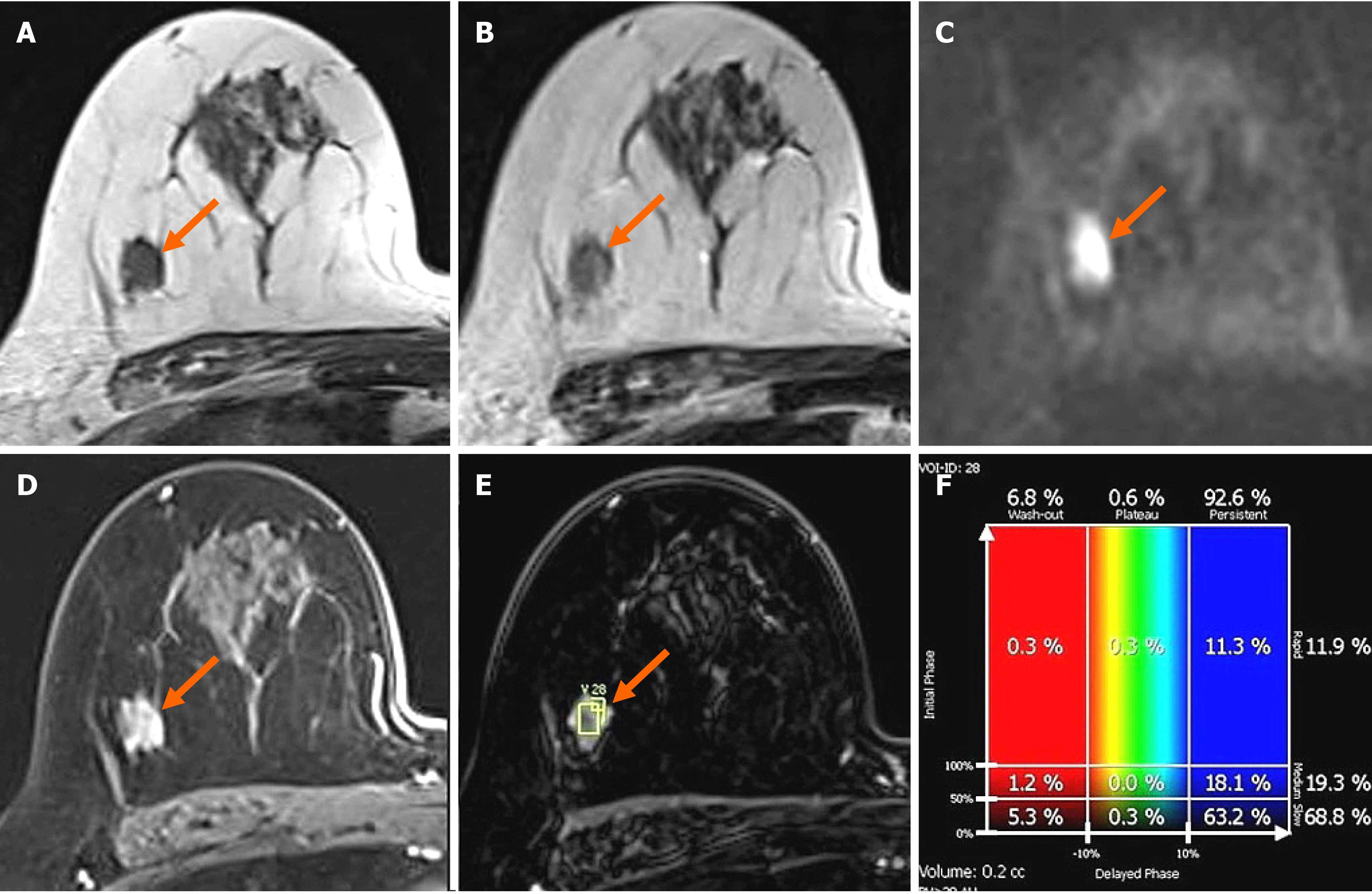
Figure 3 Breast magnetic resonance imaging showing an irregular mass in right breast.
In breast magnetic resonance imaging, the lesion showed similar T1 signal intensity (A) and slightly higher T2 signal intensity (B) compared to muscles or the breast parenchyma. The lesion showed reduced diffusivity with hyperintensity in diffusion-weighted images (C). In the contrast enhancement study, the lesion showed heterogeneous internal enhancement (D) with an initial slow and delayed persistent enhancing pattern (E and F).
- Citation: An JK, Woo JJ, Kim EK, Kwak HY. Breast adenoid cystic carcinoma arising in microglandular adenosis: A case report and review of literature. World J Clin Cases 2021; 9(25): 7579-7587
- URL: https://www.wjgnet.com/2307-8960/full/v9/i25/7579.htm
- DOI: https://dx.doi.org/10.12998/wjcc.v9.i25.7579


