Copyright
©The Author(s) 2021.
World J Clin Cases. Sep 6, 2021; 9(25): 7572-7578
Published online Sep 6, 2021. doi: 10.12998/wjcc.v9.i25.7572
Published online Sep 6, 2021. doi: 10.12998/wjcc.v9.i25.7572
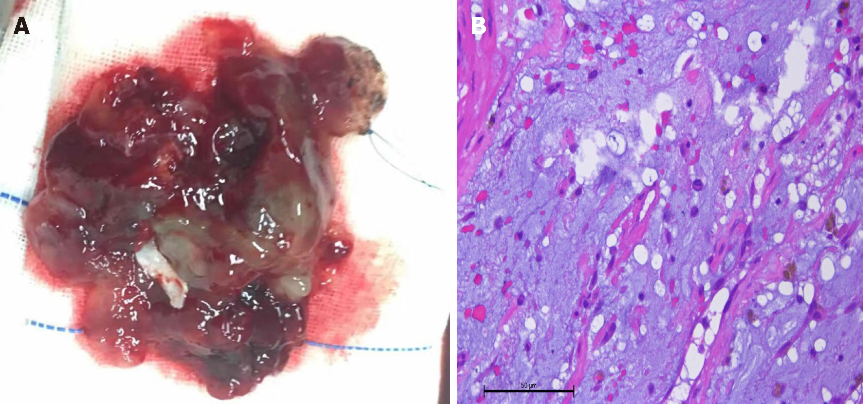
Figure 3 Appearance and microscopic display of the myxoma.
A: Gross pathology. The excised myxoma measured 6.5 cm × 6.0 cm × 5.0 cm. On macroscopic examination, the excised tumor was reddish brown jelly-like with an irregular surface and mucus; B: Histological examination of the surgically removed cardiac mass showing [hematoxylin-eosin staining (HE), scale bar: × 200] mucinous tumor cells that were scattered, shuttle-shaped or stellate, with medium amounts of cytoplasm, eosinophilic, oval nuclei, and no nuclear divisions seen in the mucinous-like matrix (HE, magnification 10 × 40).
- Citation: Chang WS, Li N, Liu H, Yin JJ, Zhang HQ. Thrombolysis and embolectomy in treatment of acute stroke as a bridge to open-heart resection of giant cardiac myxoma: A case report. World J Clin Cases 2021; 9(25): 7572-7578
- URL: https://www.wjgnet.com/2307-8960/full/v9/i25/7572.htm
- DOI: https://dx.doi.org/10.12998/wjcc.v9.i25.7572


