Copyright
©The Author(s) 2021.
World J Clin Cases. Sep 6, 2021; 9(25): 7572-7578
Published online Sep 6, 2021. doi: 10.12998/wjcc.v9.i25.7572
Published online Sep 6, 2021. doi: 10.12998/wjcc.v9.i25.7572
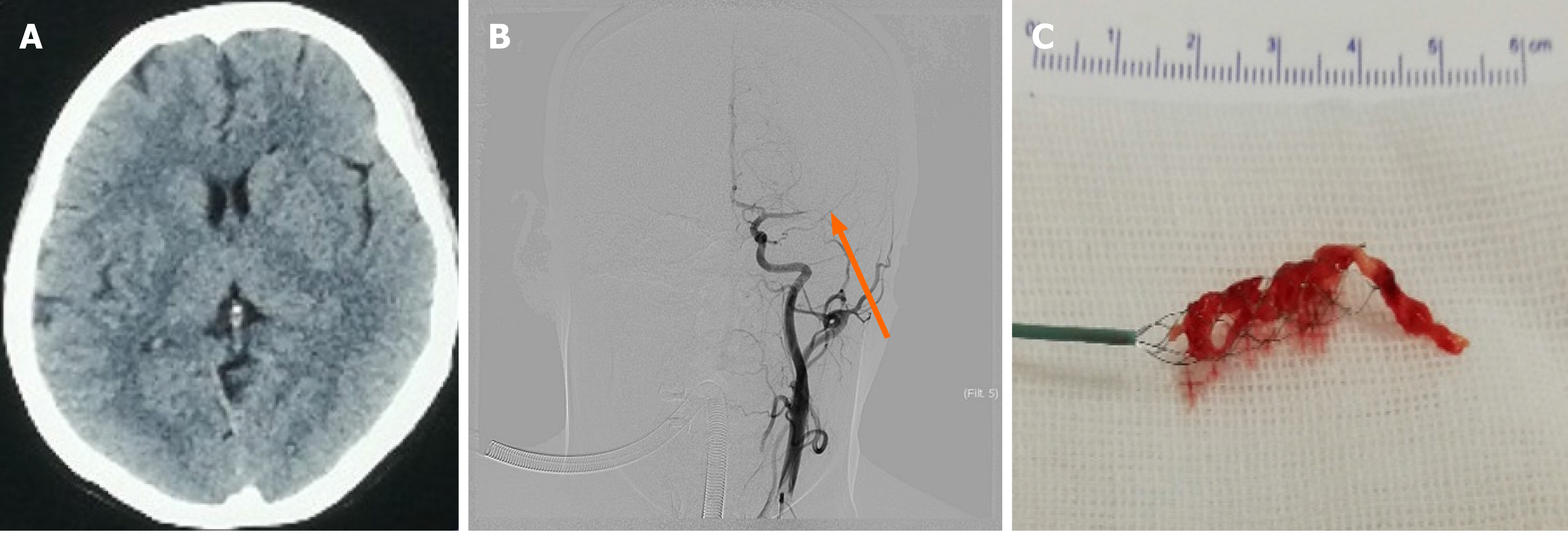
Figure 1 Head computed tomography before operation, intraoperative angiography, and stent embolectomy.
A: Head computed tomography showed no abnormalities; B: Cerebral angiography showed that the left middle cerebral artery was occluded (orange arrow); C: Thrombus removed with a stent (Solitaire FR 4.0 mm × 20 mm).
- Citation: Chang WS, Li N, Liu H, Yin JJ, Zhang HQ. Thrombolysis and embolectomy in treatment of acute stroke as a bridge to open-heart resection of giant cardiac myxoma: A case report. World J Clin Cases 2021; 9(25): 7572-7578
- URL: https://www.wjgnet.com/2307-8960/full/v9/i25/7572.htm
- DOI: https://dx.doi.org/10.12998/wjcc.v9.i25.7572


