Copyright
©The Author(s) 2021.
World J Clin Cases. Sep 6, 2021; 9(25): 7520-7526
Published online Sep 6, 2021. doi: 10.12998/wjcc.v9.i25.7520
Published online Sep 6, 2021. doi: 10.12998/wjcc.v9.i25.7520
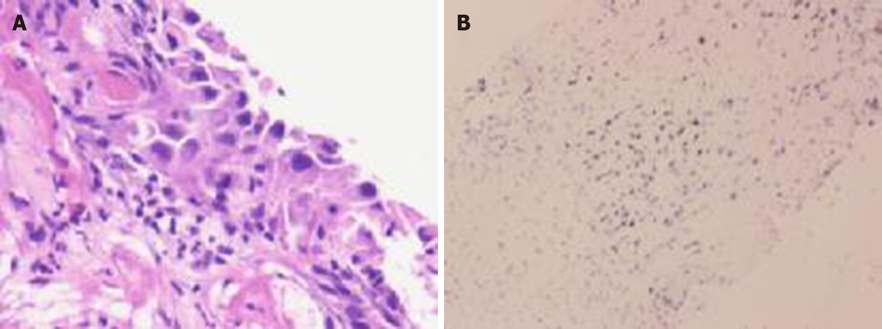
Figure 6 Histological and immunohistochemical examinations.
A: The right main bronchus mass was squamous cell carcinoma (hematoxylin and eosin staining, × 200); B: The tumor cells were positive for P63, P40, CK7, CK(Pan), Ki67, and p53 but negative for TTF-1, NapsinA, SOX2, SALL4, and CD34 (× 200).
- Citation: Jiang H, Li YQ. Coexistence of tuberculosis and squamous cell carcinoma in the right main bronchus: A case report. World J Clin Cases 2021; 9(25): 7520-7526
- URL: https://www.wjgnet.com/2307-8960/full/v9/i25/7520.htm
- DOI: https://dx.doi.org/10.12998/wjcc.v9.i25.7520


