Copyright
©The Author(s) 2021.
World J Clin Cases. Sep 6, 2021; 9(25): 7478-7483
Published online Sep 6, 2021. doi: 10.12998/wjcc.v9.i25.7478
Published online Sep 6, 2021. doi: 10.12998/wjcc.v9.i25.7478
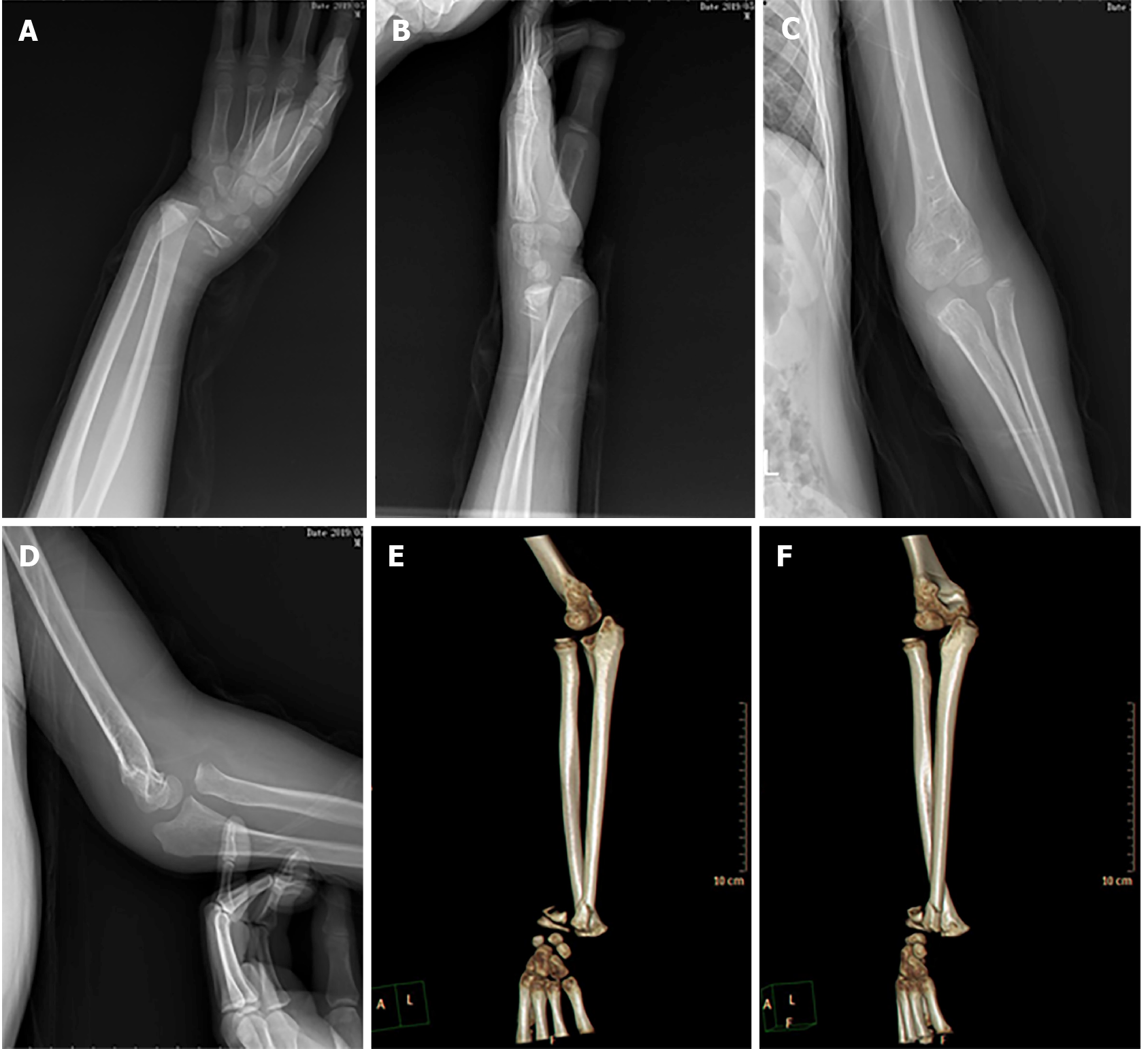
Figure 1 Preoperative imaging examination showing that the distal radius and ulna were fractured.
A: Anteroposterior X-ray of the wrist showing epiphysis injury of the distal radius with the distal fracture displaced to the radial side; B: Lateral X-ray of the wrist showing that the distal fracture was displaced to the back side; C: Anteroposterior X-ray of the elbow showing that the radial head was dislocated laterally; D: Lateral X-ray of the elbow showing that the radial head was dislocated anteriorly; E: Three-dimensional computed tomography of the forearm showing radial head dislocation with distal radial epiphysis injury; F: The relative position of the radius and ulna is crisscross.
- Citation: Jiang YK, Wang YB, Peng CG, Qu J, Wu DK. First case of forearm crisscross injury in children: A case report. World J Clin Cases 2021; 9(25): 7478-7483
- URL: https://www.wjgnet.com/2307-8960/full/v9/i25/7478.htm
- DOI: https://dx.doi.org/10.12998/wjcc.v9.i25.7478


