Copyright
©The Author(s) 2021.
World J Clin Cases. Sep 6, 2021; 9(25): 7459-7467
Published online Sep 6, 2021. doi: 10.12998/wjcc.v9.i25.7459
Published online Sep 6, 2021. doi: 10.12998/wjcc.v9.i25.7459
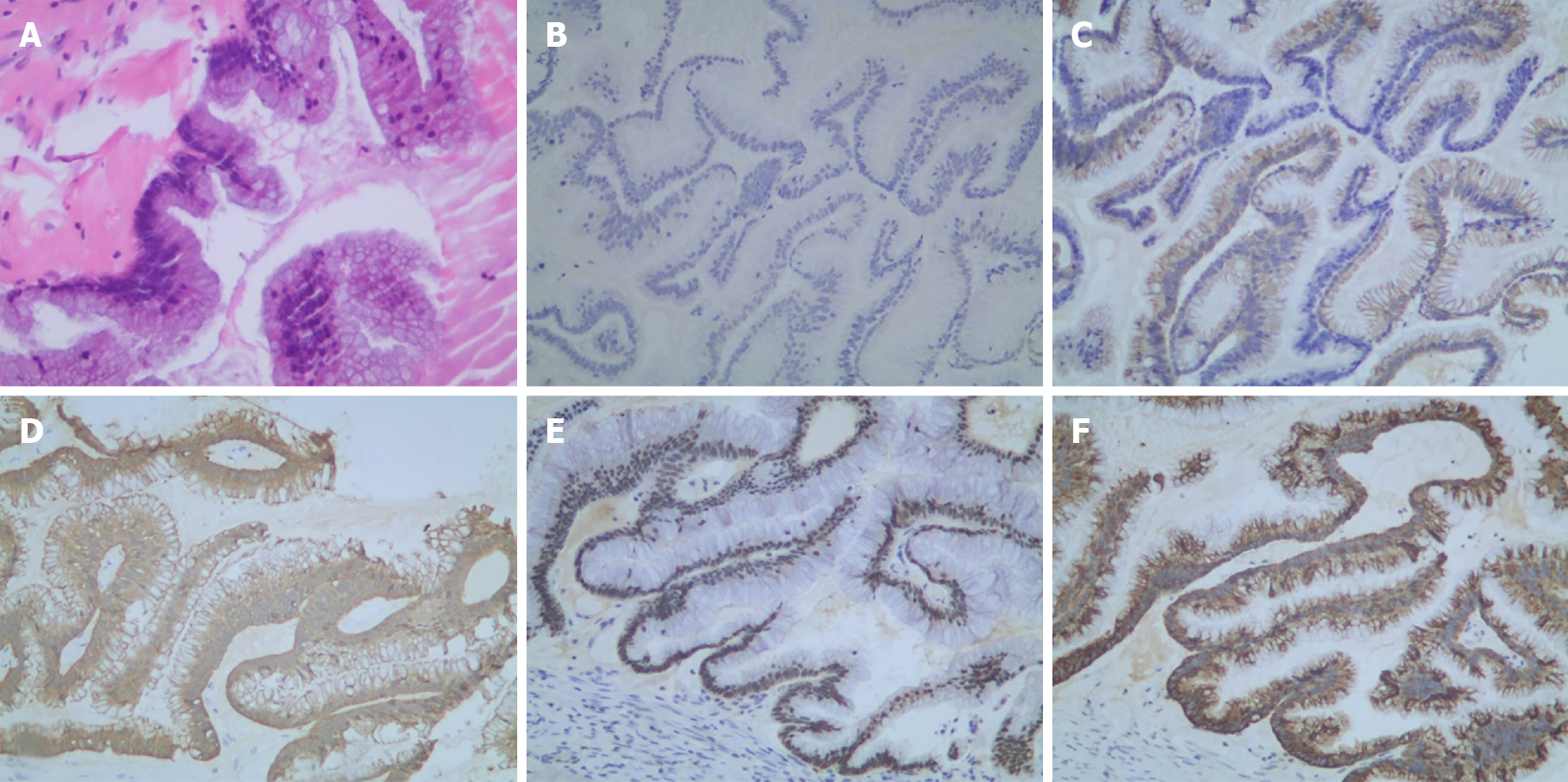
Figure 6 Histologic presentation of low-grade mucinous neoplasm.
A: Hematoxylin-eosin staining of the primary tumor; B-F: Immunohistochemical staining found that the primary tumor was CK-7(–) (B), CK-20(+) (C), Villin(+) (D), CDX-2(+) (E), and MUC-2(+) (F).
- Citation: Han XD, Zhou N, Lu YY, Xu HB, Guo J, Liang L. Pseudomyxoma peritonei originating from intestinal duplication: A case report and review of the literature. World J Clin Cases 2021; 9(25): 7459-7467
- URL: https://www.wjgnet.com/2307-8960/full/v9/i25/7459.htm
- DOI: https://dx.doi.org/10.12998/wjcc.v9.i25.7459


