Copyright
©The Author(s) 2021.
World J Clin Cases. Aug 26, 2021; 9(24): 7196-7204
Published online Aug 26, 2021. doi: 10.12998/wjcc.v9.i24.7196
Published online Aug 26, 2021. doi: 10.12998/wjcc.v9.i24.7196
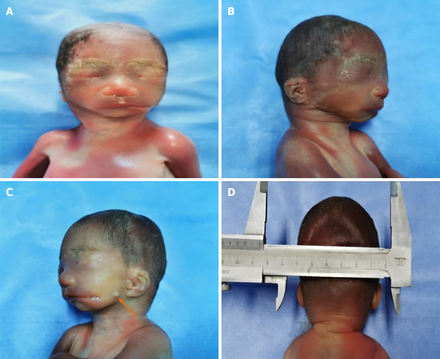
Figure 3 Pathological examination findings.
A: In the frontal view, the cleft extends from the left oral commissure to the left cheek; B and C: In the lateral view, the right face is normal, the lateral facial cleft on the left face extends to the left cheek, and a skin tag is identified anterior to the left external ear, which was not detected on ultrasonography (arrow); D: The posterior view is unremarkable. After parental counseling, the parents made an informed choice to terminate the pregnancy.
- Citation: Song WL, Ma HO, Nan Y, Li YJ, Qi N, Zhang LY, Xu X, Wang YY. Prenatal diagnosis of isolated lateral facial cleft by ultrasonography and three-dimensional printing: A case report. World J Clin Cases 2021; 9(24): 7196-7204
- URL: https://www.wjgnet.com/2307-8960/full/v9/i24/7196.htm
- DOI: https://dx.doi.org/10.12998/wjcc.v9.i24.7196


