Copyright
©The Author(s) 2021.
World J Clin Cases. Aug 26, 2021; 9(24): 7175-7180
Published online Aug 26, 2021. doi: 10.12998/wjcc.v9.i24.7175
Published online Aug 26, 2021. doi: 10.12998/wjcc.v9.i24.7175
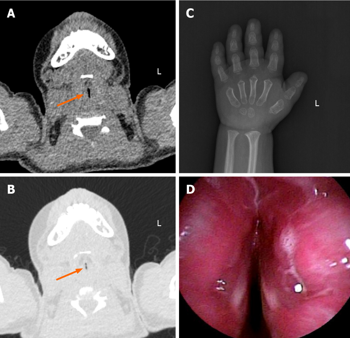
Figure 2 Representative radiographic and flexible bronchofiberscope images.
A and B: Laryngopharynx computed tomography presented trachea stenosis; C: Skeletal X-ray was taken at age 6 and revealed a delayed bone age and epiphyseal dysplasia; D: Electronic bronchoscope demonstrated severe glottic stenosis.
- Citation: Tao Y, Wei Q, Chen X, Nong GM. Geleophysic dysplasia caused by a mutation in FBN1: A case report. World J Clin Cases 2021; 9(24): 7175-7180
- URL: https://www.wjgnet.com/2307-8960/full/v9/i24/7175.htm
- DOI: https://dx.doi.org/10.12998/wjcc.v9.i24.7175


