Copyright
©The Author(s) 2021.
World J Clin Cases. Aug 26, 2021; 9(24): 7123-7132
Published online Aug 26, 2021. doi: 10.12998/wjcc.v9.i24.7123
Published online Aug 26, 2021. doi: 10.12998/wjcc.v9.i24.7123
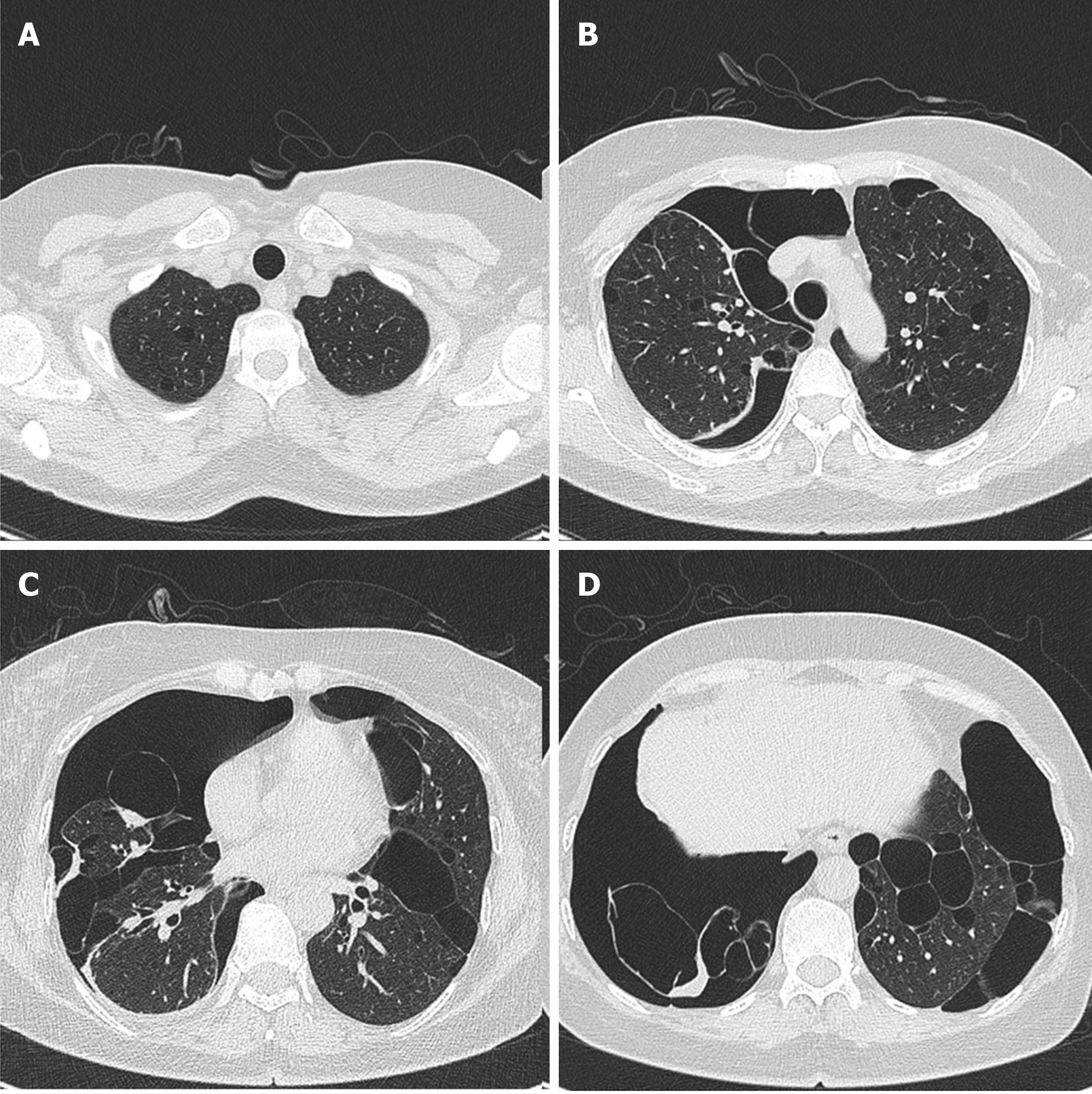
Figure 5 Axial computed tomography images.
A: The patient’s mother showed multiple thin-walled cysts in the apical segment of the right upper lobe; B: Irregular morphology cysts located in the upper-lung of both sides; C: Bilateral multiple lung cysts located at the level of the upper mediastinum; D: Right-sided pneumothorax and bilateral lung cysts located at both lung bases.
- Citation: Lu YR, Yuan Q, Liu J, Han X, Liu M, Liu QQ, Wang YG. A rare occurrence of a hereditary Birt-Hogg-Dubé syndrome: A case report. World J Clin Cases 2021; 9(24): 7123-7132
- URL: https://www.wjgnet.com/2307-8960/full/v9/i24/7123.htm
- DOI: https://dx.doi.org/10.12998/wjcc.v9.i24.7123


