Copyright
©The Author(s) 2021.
World J Clin Cases. Aug 26, 2021; 9(24): 7110-7116
Published online Aug 26, 2021. doi: 10.12998/wjcc.v9.i24.7110
Published online Aug 26, 2021. doi: 10.12998/wjcc.v9.i24.7110
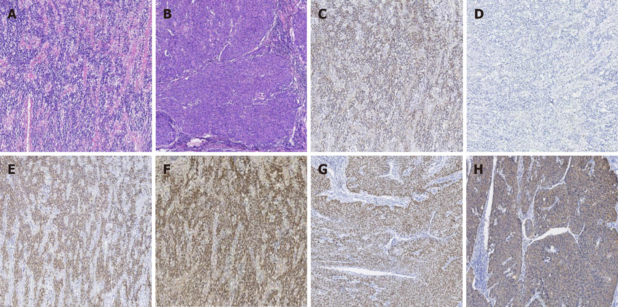
Figure 2 Pathology results of extramedullary plasmacytoma.
A: Hematoxylin-eosin (HE) staining of extramedullary plasmacytoma. The tumor cells were diffusely distributed. Pathological spindle division and Russell body were observed; B: HE staining of cervical squamous cell carcinoma. Hyperplasia of epithelioid cell nests, infiltrating growth pathological fission, and intercellular Bridges were easily observed; C: Immunohistochemistry staining of Kappa, which was diffusely positive in Plasma tumor cells; D: Immunohistochemistry staining of Lambda, which was diffusely negative in Plasma tumor cells; E: Immunohistochemistry staining of CD38, which was diffusely positive in Plasma tumor cells; F: Immunohistochemistry staining of CD138, which was diffusely positive in Plasma tumor cells; G: Immunohistochemistry staining of p40, which was diffusely positive in cervical squamous cancer cells; H: Immunohistochemistry staining of CK5/6, which was diffusely positive in cervical squamous cancer cells. Magnification: A-H × 40.
- Citation: Zhang QY, Li TC, Lin J, He LL, Liu XY. Coexistence of cervical extramedullary plasmacytoma and squamous cell carcinoma: A case report. World J Clin Cases 2021; 9(24): 7110-7116
- URL: https://www.wjgnet.com/2307-8960/full/v9/i24/7110.htm
- DOI: https://dx.doi.org/10.12998/wjcc.v9.i24.7110


