Copyright
©The Author(s) 2021.
World J Clin Cases. Aug 16, 2021; 9(23): 6886-6899
Published online Aug 16, 2021. doi: 10.12998/wjcc.v9.i23.6886
Published online Aug 16, 2021. doi: 10.12998/wjcc.v9.i23.6886
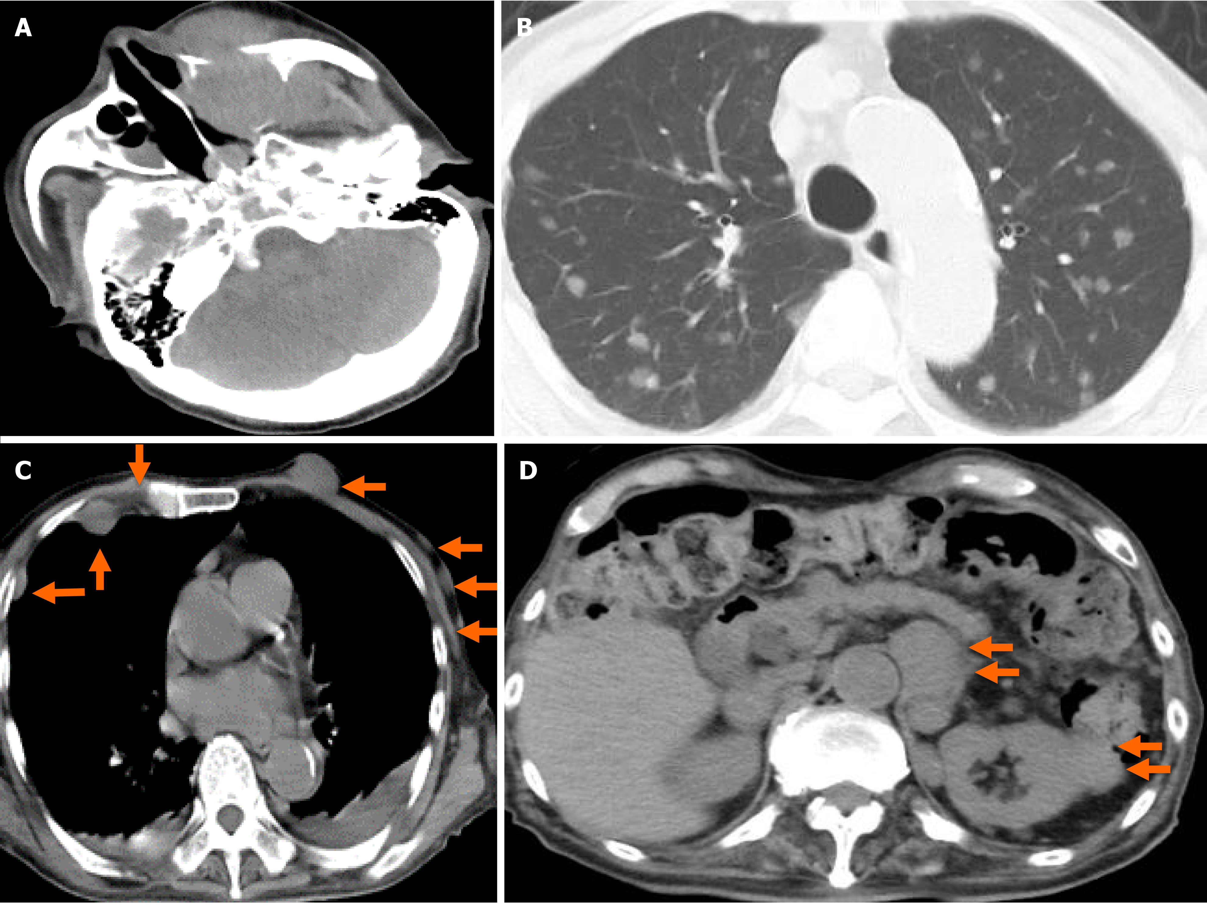
Figure 4 A plain computed tomography scan on day 62.
A: The 4 cm × 3.5 cm × 4 cm sized left maxillary sinus was completely filled with mass, and it was oppressed, involved the orbital base, destroyed the anterior, internal, and external walls, and protuberated outside; B: Chest. Pulmonary window setting. Multiple tumors (0.5-1.5 cm in size) were confirmed in both lungs; C: Chest. Mediastinal window. Multiple subcutaneous tumors and chest wall tumors were confirmed (orange arrows); and D: Abdomen. An abdominal paraaortic lymph node swelling (3 cm in size) and a left kidney nodule (1-2 cm in size) were confirmed (orange arrows).
- Citation: Usuda D, Izumida T, Terada N, Sangen R, Higashikawa T, Sekiguchi S, Tanaka R, Suzuki M, Hotchi Y, Shimozawa S, Tokunaga S, Osugi I, Katou R, Ito S, Asako S, Takagi Y, Mishima K, Kondo A, Mizuno K, Takami H, Komatsu T, Oba J, Nomura T, Sugita M, Kasamaki Y. Diffuse large B cell lymphoma originating from the maxillary sinus with skin metastases: A case report and review of literature. World J Clin Cases 2021; 9(23): 6886-6899
- URL: https://www.wjgnet.com/2307-8960/full/v9/i23/6886.htm
- DOI: https://dx.doi.org/10.12998/wjcc.v9.i23.6886


