Copyright
©The Author(s) 2021.
World J Clin Cases. Aug 16, 2021; 9(23): 6886-6899
Published online Aug 16, 2021. doi: 10.12998/wjcc.v9.i23.6886
Published online Aug 16, 2021. doi: 10.12998/wjcc.v9.i23.6886
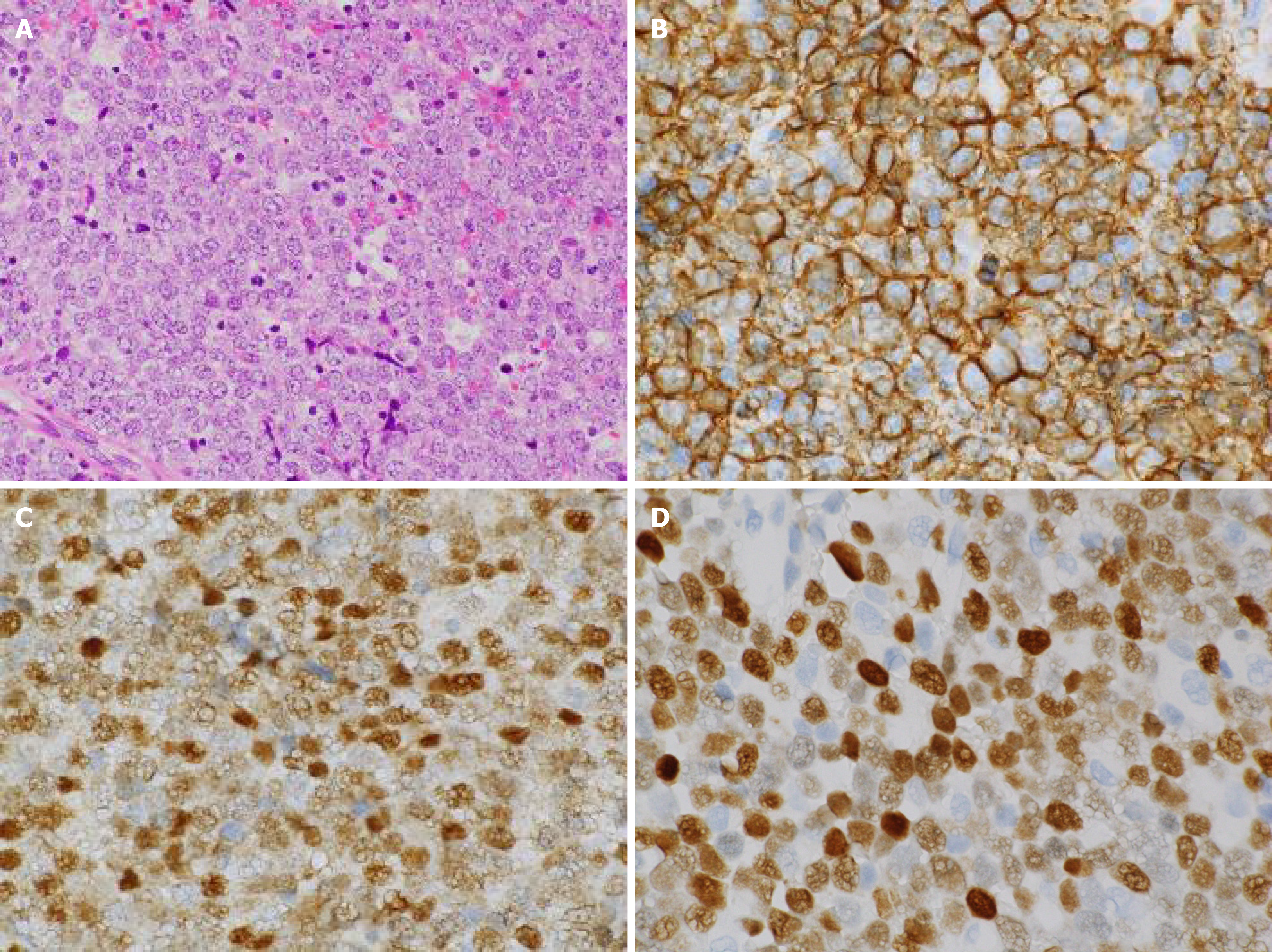
Figure 3 Histopathological images of the biopsy specimen from the left maxillary sinus mass taken on day 50.
A: Aggregates of large atypical lymphocytes with irregular nuclei having uneven chromatin and small to large nucleoli are evident in a necrotic background. Some cells show multilobulated nuclei. Mitosis and apoptotic bodies are conspicuous. Hematoxylin and eosin staining (× 200 magnification); B: Cluster of differentiation 20-positive cells were confirmed through immunohistochemical staining (× 400 magnification); C: Bcl-6-positive cells were confirmed through immunohistochemical staining (× 400 magnification); and D: MUM1-positive cells were confirmed through immunohistochemical staining (× 400 magnification).
- Citation: Usuda D, Izumida T, Terada N, Sangen R, Higashikawa T, Sekiguchi S, Tanaka R, Suzuki M, Hotchi Y, Shimozawa S, Tokunaga S, Osugi I, Katou R, Ito S, Asako S, Takagi Y, Mishima K, Kondo A, Mizuno K, Takami H, Komatsu T, Oba J, Nomura T, Sugita M, Kasamaki Y. Diffuse large B cell lymphoma originating from the maxillary sinus with skin metastases: A case report and review of literature. World J Clin Cases 2021; 9(23): 6886-6899
- URL: https://www.wjgnet.com/2307-8960/full/v9/i23/6886.htm
- DOI: https://dx.doi.org/10.12998/wjcc.v9.i23.6886


