Copyright
©The Author(s) 2021.
World J Clin Cases. Aug 16, 2021; 9(23): 6781-6788
Published online Aug 16, 2021. doi: 10.12998/wjcc.v9.i23.6781
Published online Aug 16, 2021. doi: 10.12998/wjcc.v9.i23.6781
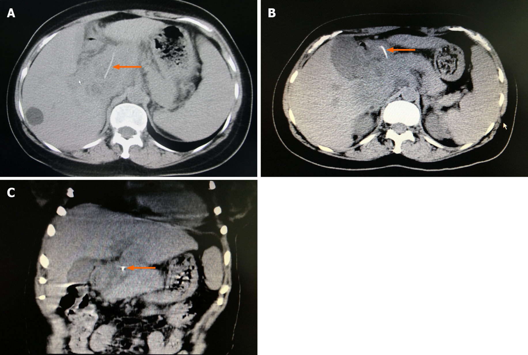
Figure 3 Computed tomography scan of the liver, gallbladder, and spleen.
A and B: An irregular soft tissue density shadow with poorly defined borders (orange arrow) is seen above the hilum, caudate lobe, and pancreatic head. A more hypodense focus is seen inside (orange arrow), measuring about 7.8 cm × 6.0 cm × 5.0 cm. A dense shadow of about 3.7 cm in length is seen inside the lesion, the anterior end of which is located in the gastric cavity; C: A foreign body (fishbone) is seen in the upper abdomen (orange arrow) and was considered to have penetrated the gastric wall to the hepatic hilum, with an abscess having formed above the caudal lobe and pancreatic head.
- Citation: Pan W, Lin LJ, Meng ZW, Cai XR, Chen YL. Hepatic abscess caused by esophageal foreign body misdiagnosed as cystadenocarcinoma by magnetic resonance imaging: A case report. World J Clin Cases 2021; 9(23): 6781-6788
- URL: https://www.wjgnet.com/2307-8960/full/v9/i23/6781.htm
- DOI: https://dx.doi.org/10.12998/wjcc.v9.i23.6781


