Copyright
©The Author(s) 2021.
World J Clin Cases. Aug 16, 2021; 9(23): 6781-6788
Published online Aug 16, 2021. doi: 10.12998/wjcc.v9.i23.6781
Published online Aug 16, 2021. doi: 10.12998/wjcc.v9.i23.6781
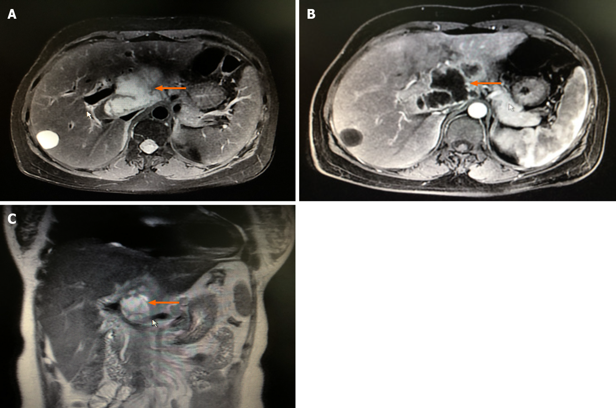
Figure 2 Enhanced magnetic resonance imaging of the liver.
A and C: A space-occupying lesion (orange arrows) is seen axially (A) and coronally (C) in the caudal lobe of the liver, with unclear borders and irregular shape, about 7.6 cm × 4.4 cm × 5.0 cm in size, slightly low signal on T1WI and slightly high signal on T2WI, and inhomogeneous signal; B: The edge of the tumor is enhanced in the arterial phase, with multiple small and disorganized vascular shadows present within the lesion. Considering the possibility of cystadenocarcinoma, it was considered in differential diagnosis of liver abscess.
- Citation: Pan W, Lin LJ, Meng ZW, Cai XR, Chen YL. Hepatic abscess caused by esophageal foreign body misdiagnosed as cystadenocarcinoma by magnetic resonance imaging: A case report. World J Clin Cases 2021; 9(23): 6781-6788
- URL: https://www.wjgnet.com/2307-8960/full/v9/i23/6781.htm
- DOI: https://dx.doi.org/10.12998/wjcc.v9.i23.6781


