Copyright
©The Author(s) 2021.
World J Clin Cases. Aug 6, 2021; 9(22): 6566-6574
Published online Aug 6, 2021. doi: 10.12998/wjcc.v9.i22.6566
Published online Aug 6, 2021. doi: 10.12998/wjcc.v9.i22.6566
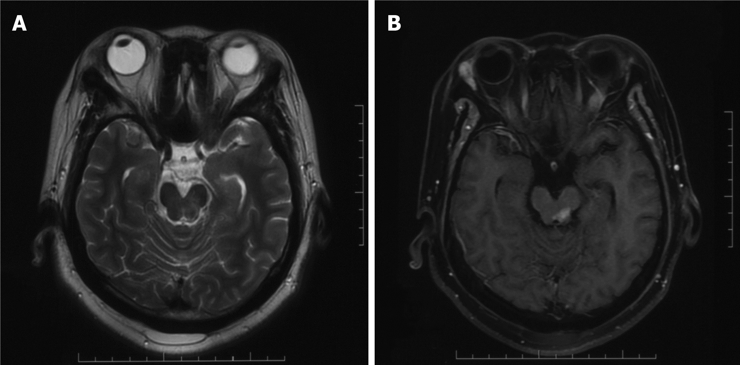
Figure 5 Magnetic resonance imaging reexamination after 20 Gy radiotherapy.
A: T2-weighted image showing mixed signal in the left midbrain; B: Contrast-enhanced magnetic resonance imaging showing residual lesion.
- Citation: Zhao YR, Hu RH, Wu R, Xu JK. Primary mucosa-associated lymphoid tissue lymphoma in the midbrain: A case report. World J Clin Cases 2021; 9(22): 6566-6574
- URL: https://www.wjgnet.com/2307-8960/full/v9/i22/6566.htm
- DOI: https://dx.doi.org/10.12998/wjcc.v9.i22.6566


