Copyright
©The Author(s) 2021.
World J Clin Cases. Aug 6, 2021; 9(22): 6493-6500
Published online Aug 6, 2021. doi: 10.12998/wjcc.v9.i22.6493
Published online Aug 6, 2021. doi: 10.12998/wjcc.v9.i22.6493
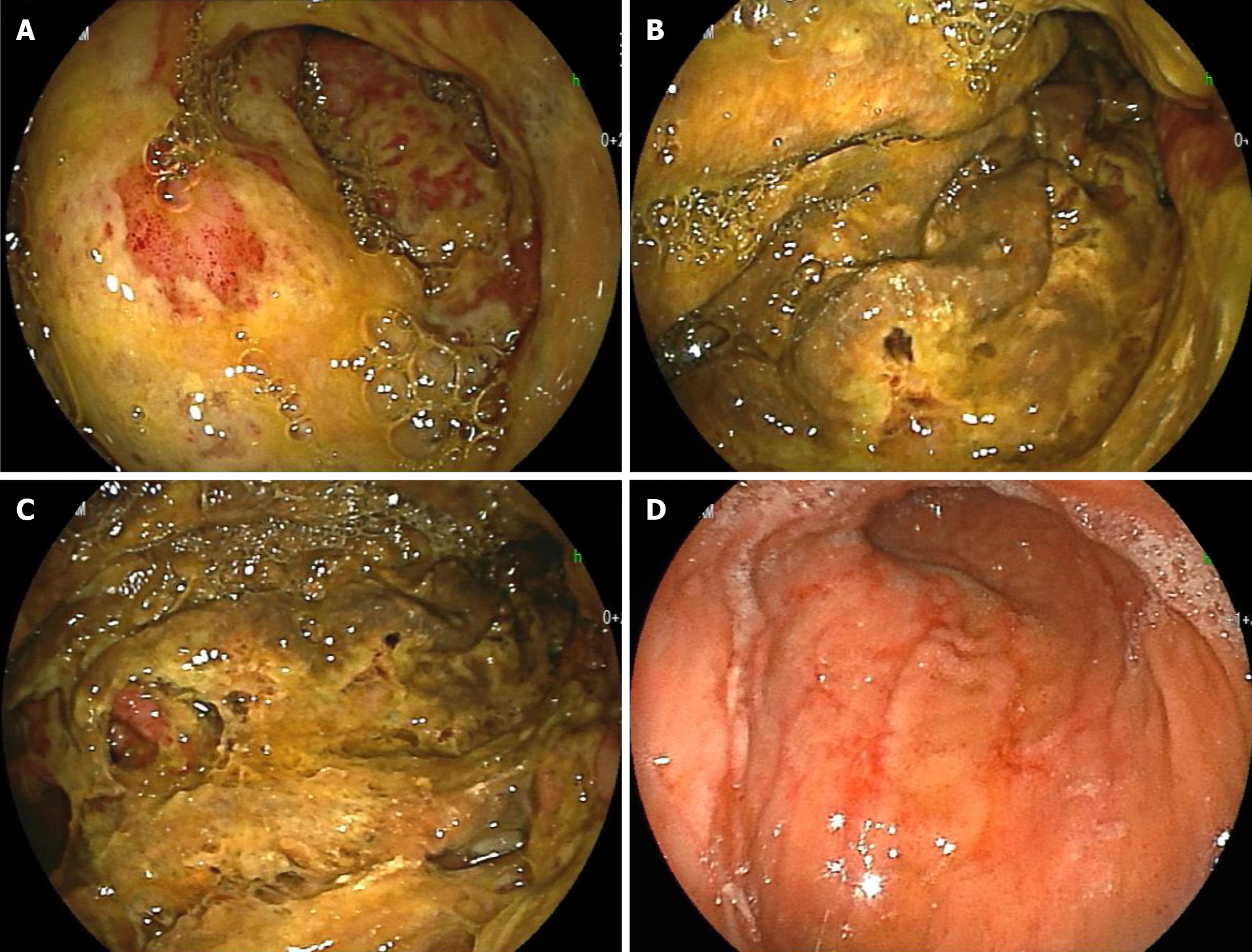
Figure 3 Esophagogastroduodenoscopy.
A-C: Marked thickening of the gastric wall in the corpus of the stomach and yellow-green pseudomembrane-like tissue covering the superficial mucosa were observed (day 14); D: The above abnormal findings were improved, and linear redness, erosion and ulcerative mucosal changes were observed (day 29).
- Citation: Saito M, Morioka M, Izumiyama K, Mori A, Ogasawara R, Kondo T, Miyajima T, Yokoyama E, Tanikawa S. Phlegmonous gastritis developed during chemotherapy for acute lymphocytic leukemia: A case report. World J Clin Cases 2021; 9(22): 6493-6500
- URL: https://www.wjgnet.com/2307-8960/full/v9/i22/6493.htm
- DOI: https://dx.doi.org/10.12998/wjcc.v9.i22.6493


