Copyright
©The Author(s) 2021.
World J Clin Cases. Aug 6, 2021; 9(22): 6469-6477
Published online Aug 6, 2021. doi: 10.12998/wjcc.v9.i22.6469
Published online Aug 6, 2021. doi: 10.12998/wjcc.v9.i22.6469
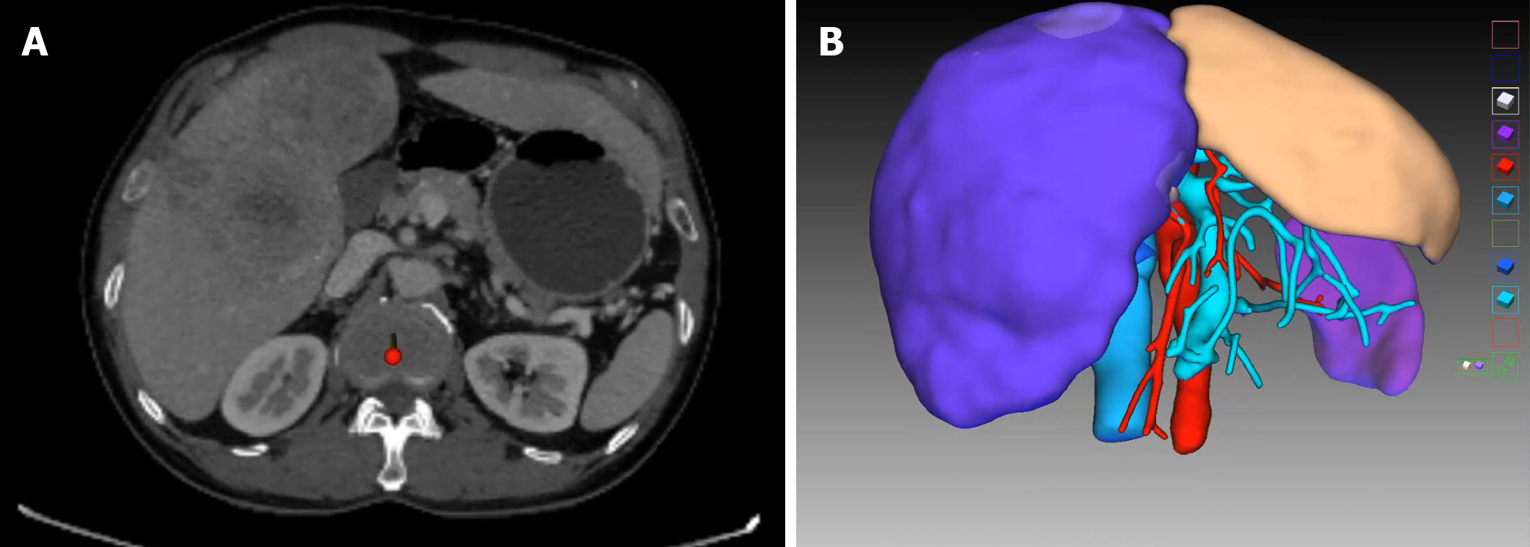
Figure 1 Computed tomography result and three-dimensional reconstruction of the patient at first presentation.
A: Venous phase contrast-enhanced computed tomography shows huge space-occupying lesions in the left medial lobe and right liver, and the left lateral lobe is normal; B: Virtual resection on the three-dimensional liver model.
- Citation: Zhang JJ, Wang ZX, Niu JX, Zhang M, An N, Li PF, Zheng WH. Successful totally laparoscopic right trihepatectomy following conversion therapy for hepatocellular carcinoma: A case report. World J Clin Cases 2021; 9(22): 6469-6477
- URL: https://www.wjgnet.com/2307-8960/full/v9/i22/6469.htm
- DOI: https://dx.doi.org/10.12998/wjcc.v9.i22.6469


