Copyright
©The Author(s) 2021.
World J Clin Cases. Aug 6, 2021; 9(22): 6435-6442
Published online Aug 6, 2021. doi: 10.12998/wjcc.v9.i22.6435
Published online Aug 6, 2021. doi: 10.12998/wjcc.v9.i22.6435
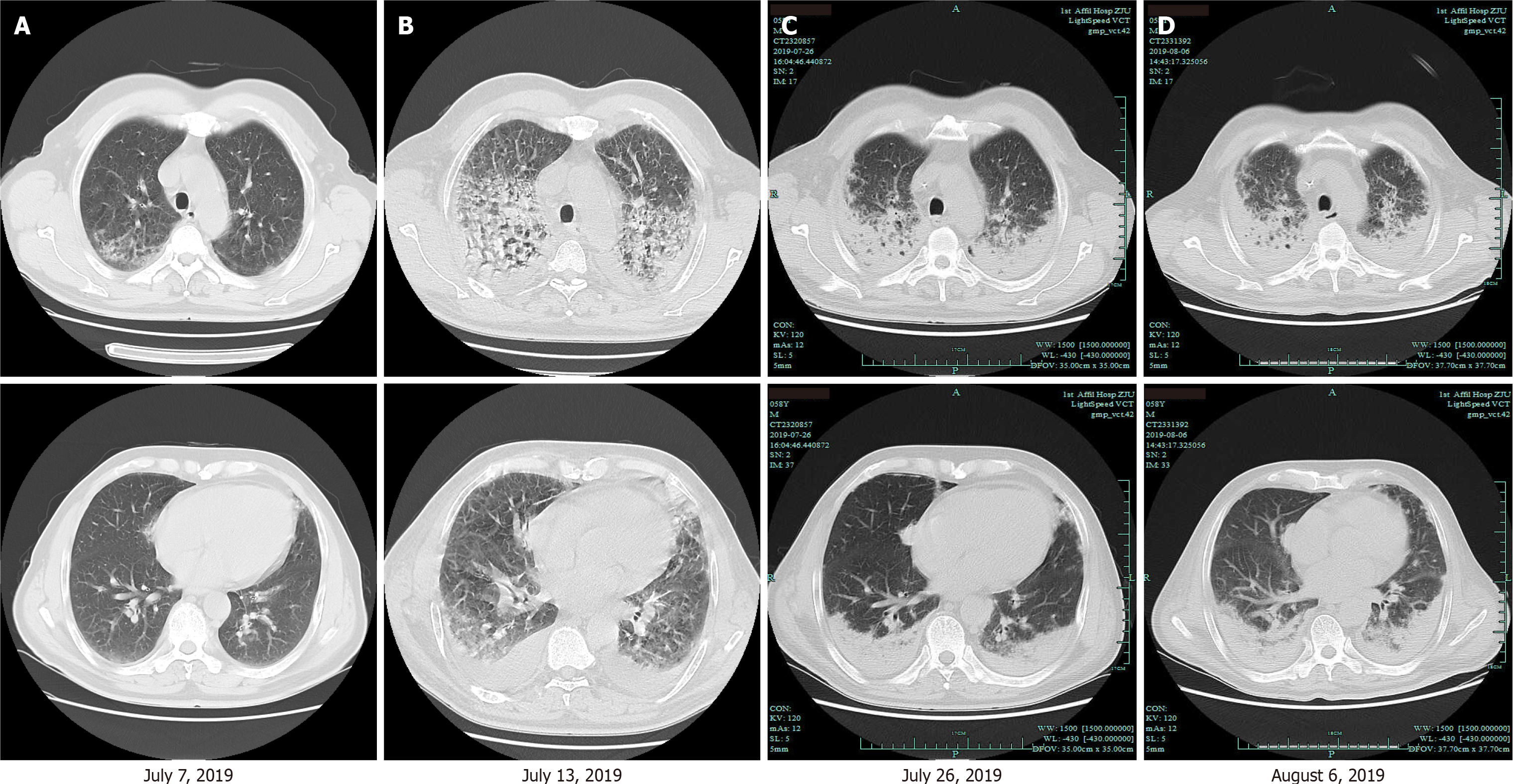
Figure 1 Serial computed tomography scans of the chest.
A: The computed tomography (CT) scan on July 7, 2019 showed bilateral ground-glass infiltrates in both upper lungs and lesions in the lingular segment of the left upper lobe and right middle lobe; B: The CT scan on July 13, 2019 showed irregular linear opacities and diffuse ground-glass opacities in both upper lungs and the bilateral pleural effusion. The lesions improved after meropenem plus moxifloxacin treatment; C: The CT scan on July 26, 2019 showed gradual bilateral consolidation in upper lobes and inferior lobes after imipenem-cilastatin plus caspofungin plus oral oseltamivir treatment; D: The CT scan on August 6, 2019 showed improvement in the bilateral consolidation after cefoperazone/sulbactam plus tigecycline treatment.
- Citation: Xu XL, Zhang RH, Wang YH, Zhou JY. Manifestation of severe pneumonia in anti-PL-7 antisynthetase syndrome and B cell lymphoma: A case report. World J Clin Cases 2021; 9(22): 6435-6442
- URL: https://www.wjgnet.com/2307-8960/full/v9/i22/6435.htm
- DOI: https://dx.doi.org/10.12998/wjcc.v9.i22.6435


