Copyright
©The Author(s) 2021.
World J Clin Cases. Aug 6, 2021; 9(22): 6428-6434
Published online Aug 6, 2021. doi: 10.12998/wjcc.v9.i22.6428
Published online Aug 6, 2021. doi: 10.12998/wjcc.v9.i22.6428
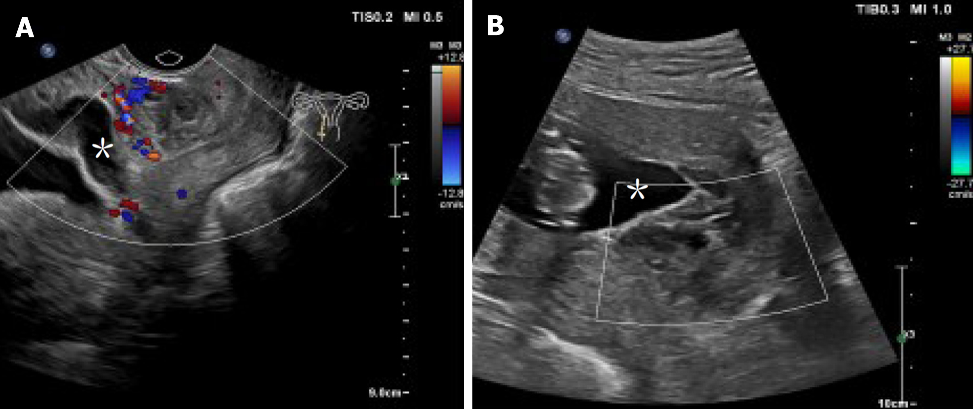
Figure 3 Sequential views of an intrauterine pregnancy and the blood supply at the site of implantation of an ectopic pregnancy on ultrasound after intervention.
A: Transvaginal color Doppler ultrasound showing the normal growth of the intrauterine pregnancy (*) and a hematoma-rich blood supply at the site of implantation of the ectopic pregnancy at 10 wk gestation; B: Transabdominal color Doppler ultrasound showing the normally growing embryo (*) and no blood supply at the site of implantation of the ectopic pregnancy at 13 wk gestation.
- Citation: Chen ZY, Zhou Y, Qian Y, Luo JM, Huang XF, Zhang XM. Management of heterotopic cesarean scar pregnancy with preservation of intrauterine pregnancy: A case report. World J Clin Cases 2021; 9(22): 6428-6434
- URL: https://www.wjgnet.com/2307-8960/full/v9/i22/6428.htm
- DOI: https://dx.doi.org/10.12998/wjcc.v9.i22.6428


