Copyright
©The Author(s) 2021.
World J Clin Cases. Aug 6, 2021; 9(22): 6428-6434
Published online Aug 6, 2021. doi: 10.12998/wjcc.v9.i22.6428
Published online Aug 6, 2021. doi: 10.12998/wjcc.v9.i22.6428
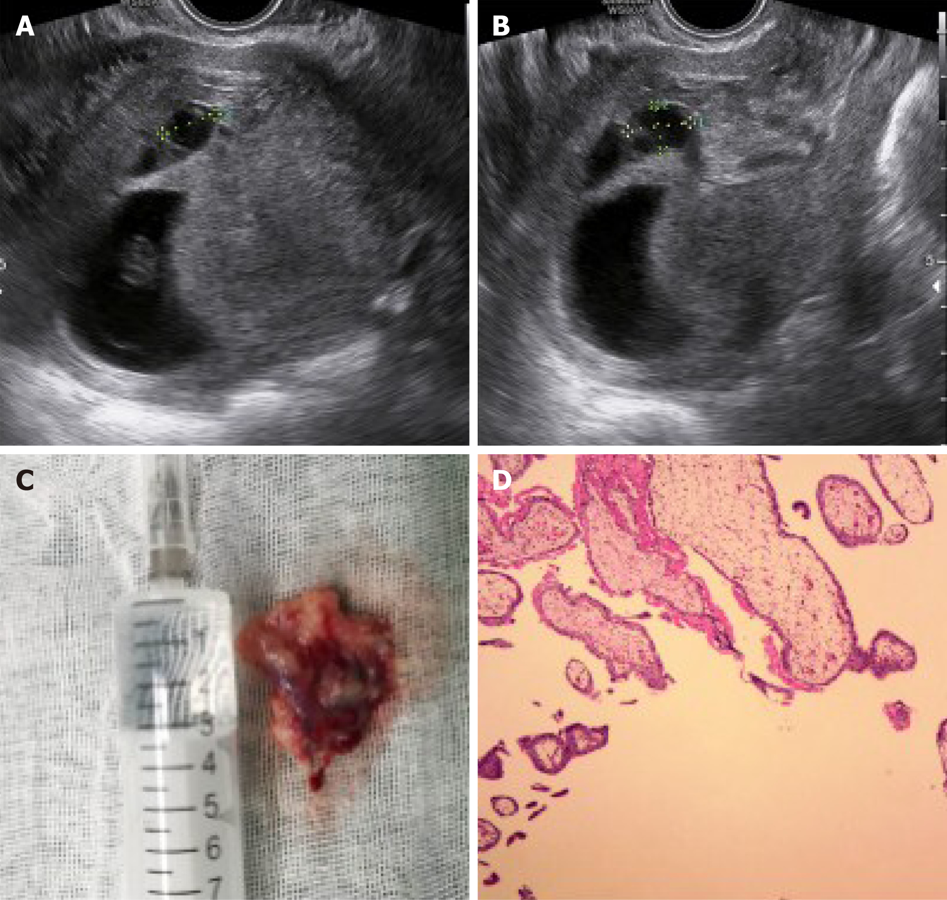
Figure 2 Ultrasound images of a heterotopic cesarean scar pregnancy after intervention.
A and B: Transvaginal ultrasound showing normal growth of the intrauterine pregnancy (gestational sac 1) in the uterine fundus and the decreased size of the ectopic pregnancy sac (gestational sac 2) in the lower uterine segment 2 d after selective embryo aspiration; C: Gestational tissue removed from the lower segment of the uterus by suction and curettage, the size marked with a 10 mL syringe; D: Histological examination of the removed specimen confirming the presence of trophoblastic tissue (hematoxylin and eosin stain, × 50).
- Citation: Chen ZY, Zhou Y, Qian Y, Luo JM, Huang XF, Zhang XM. Management of heterotopic cesarean scar pregnancy with preservation of intrauterine pregnancy: A case report. World J Clin Cases 2021; 9(22): 6428-6434
- URL: https://www.wjgnet.com/2307-8960/full/v9/i22/6428.htm
- DOI: https://dx.doi.org/10.12998/wjcc.v9.i22.6428


