Copyright
©The Author(s) 2021.
World J Clin Cases. Aug 6, 2021; 9(22): 6418-6427
Published online Aug 6, 2021. doi: 10.12998/wjcc.v9.i22.6418
Published online Aug 6, 2021. doi: 10.12998/wjcc.v9.i22.6418
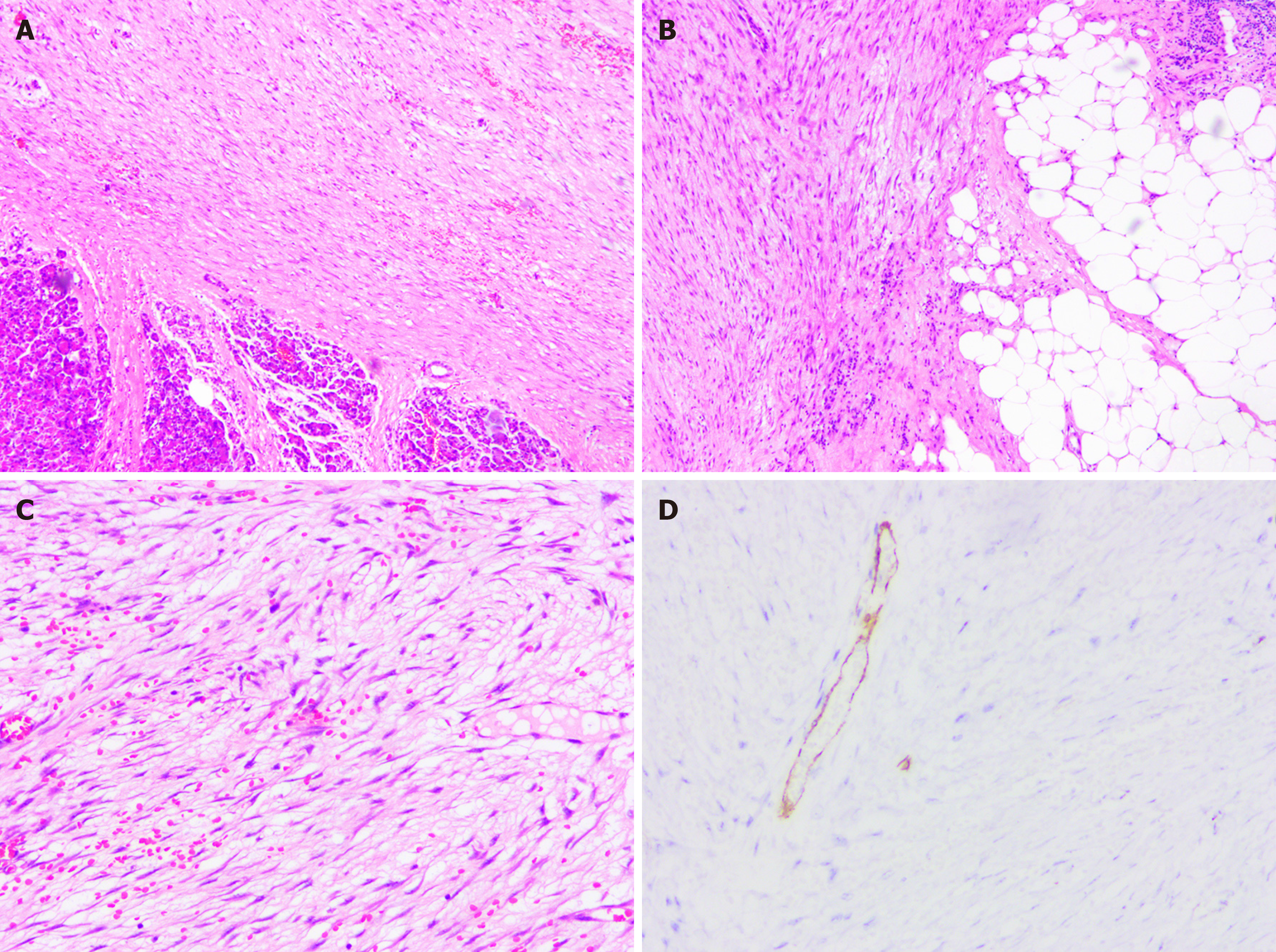
Figure 2 The pathological findings of the resected specimen revealed inflammatory myofibroblastic tumor.
A-C: Histological images showed mixed components of dense myofibroblastic tissues and few inflammatory cells, with neoplastic cells infiltrating the surrounding fat tissue (hematoxylin and eosin staining); D: Immunohistochemical studies showed positivity for smooth muscle actin.
- Citation: Chen ZT, Lin YX, Li MX, Zhang T, Wan DL, Lin SZ. Inflammatory myofibroblastic tumor of the pancreatic neck: A case report and review of literature. World J Clin Cases 2021; 9(22): 6418-6427
- URL: https://www.wjgnet.com/2307-8960/full/v9/i22/6418.htm
- DOI: https://dx.doi.org/10.12998/wjcc.v9.i22.6418


