Copyright
©The Author(s) 2021.
World J Clin Cases. Aug 6, 2021; 9(22): 6380-6387
Published online Aug 6, 2021. doi: 10.12998/wjcc.v9.i22.6380
Published online Aug 6, 2021. doi: 10.12998/wjcc.v9.i22.6380
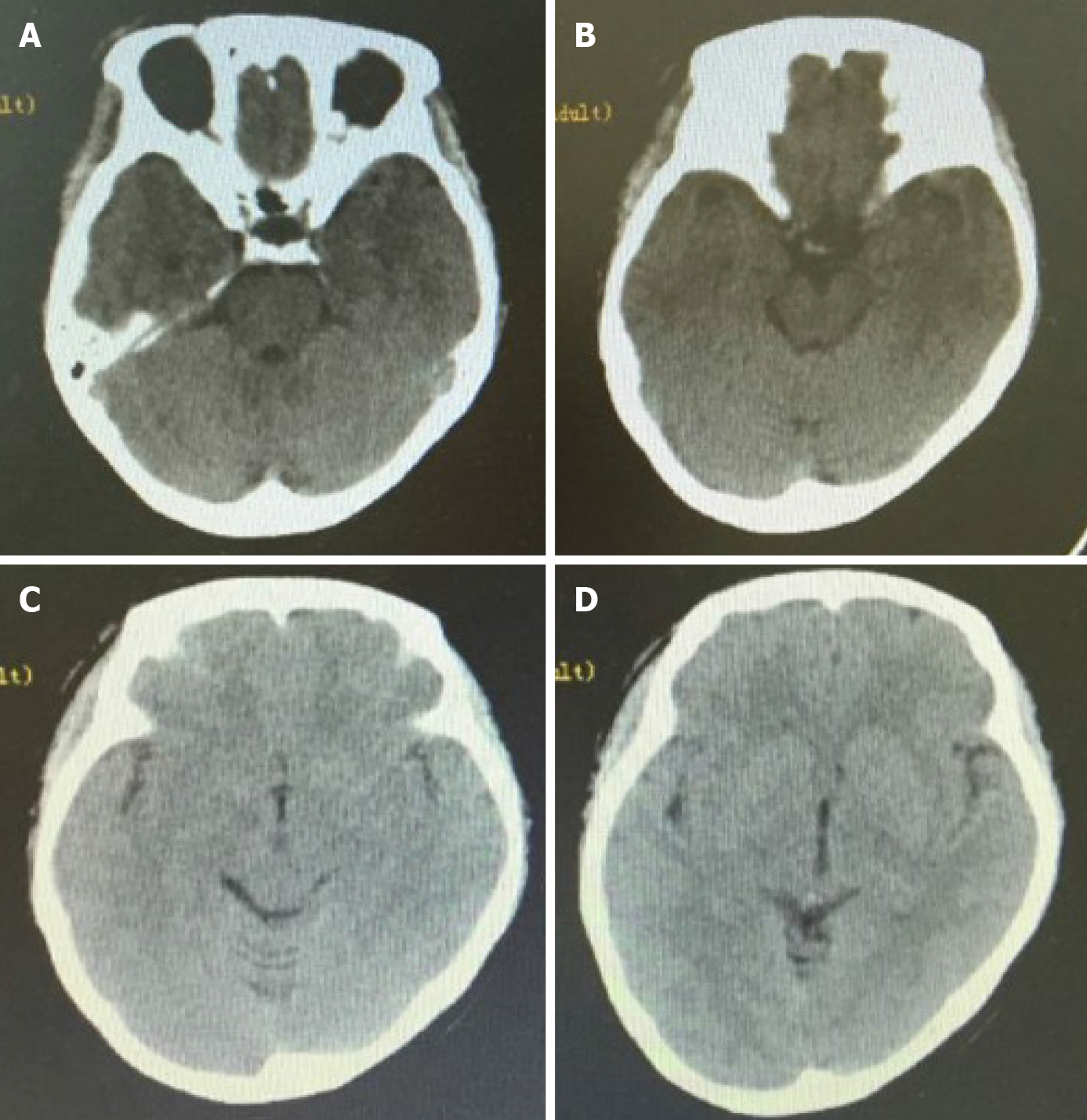
Figure 1 First head computed tomography images.
A: No abnormal density lesion was found near the parasellar region; B: No abnormal density lesion or subarachnoid hemorrhage was observed in the suprasellar cistern and annular cistern; C and D: No obvious subarachnoid hemorrhage was observed in any sulci or cistern.
- Citation: Zhao L, Zhao SQ, Tang XP. Ruptured intracranial aneurysm presenting as cerebral circulation insufficiency: A case report. World J Clin Cases 2021; 9(22): 6380-6387
- URL: https://www.wjgnet.com/2307-8960/full/v9/i22/6380.htm
- DOI: https://dx.doi.org/10.12998/wjcc.v9.i22.6380


