Copyright
©The Author(s) 2021.
World J Clin Cases. Aug 6, 2021; 9(22): 6254-6267
Published online Aug 6, 2021. doi: 10.12998/wjcc.v9.i22.6254
Published online Aug 6, 2021. doi: 10.12998/wjcc.v9.i22.6254
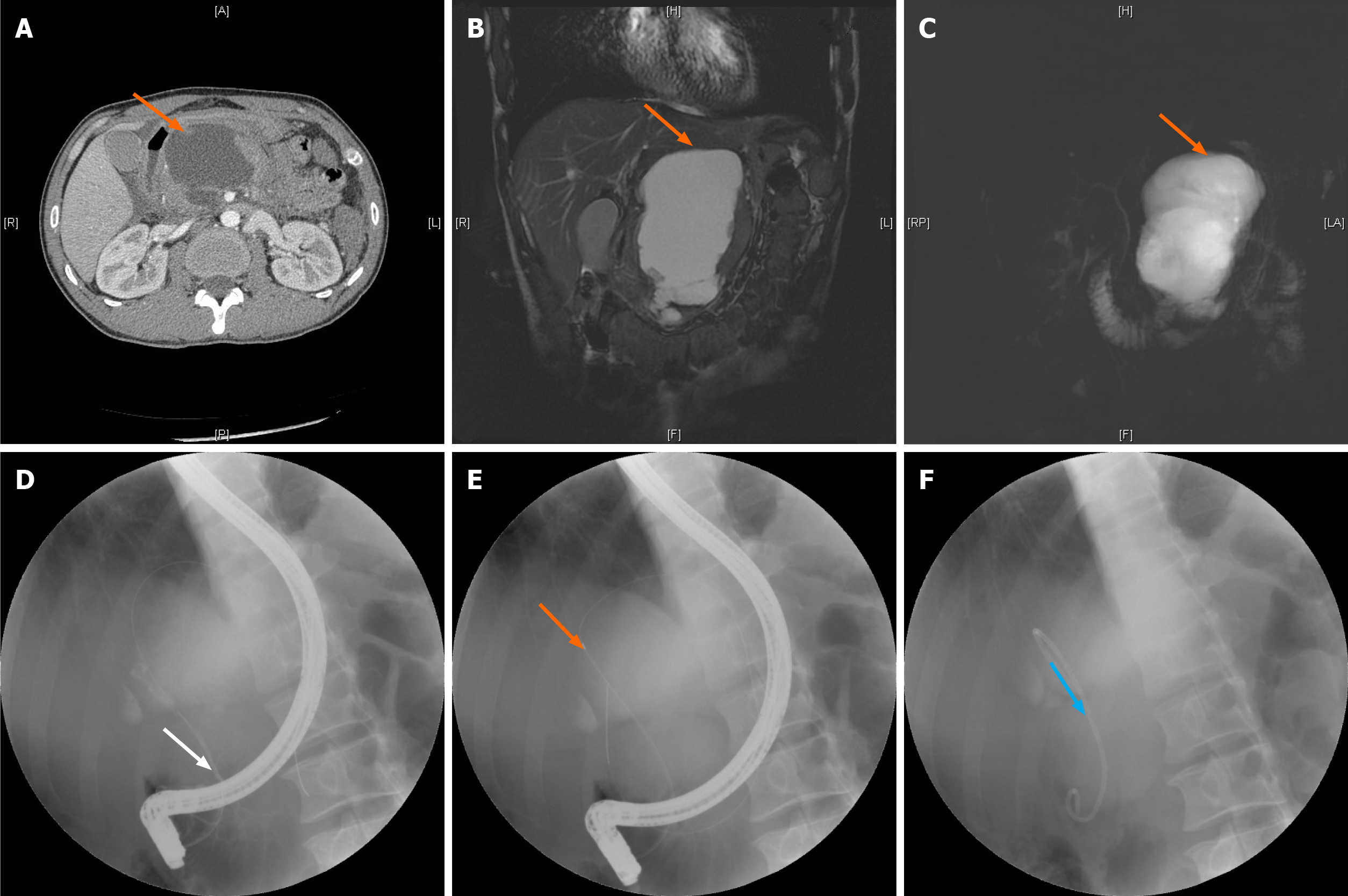
Figure 1 Process of pancreatic duct stenting for pancreatic pseudocysts.
A: Location of the pancreatic pseudocyst shown by computed tomography; B and C: Location of the pancreatic pseudocyst shown by magnetic resonance cholangiopancreatography (orange arrow) using the T2 weighted image sequence (B) and balanced turbo field echo sequence (C); D: Successful duodenal intubation followed by a guide wire inserted into the pancreatic duct (white arrow); E: Pancreatic duct and cyst shown by imaging (orange arrow); F: Pancreatic duct stenting (blue arrow).
- Citation: He YG, Li J, Peng XH, Wu J, Xie MX, Tang YC, Zheng L, Huang XB. Sequential therapy with combined trans-papillary endoscopic naso-pancreatic and endoscopic retrograde pancreatic drainage for pancreatic pseudocysts. World J Clin Cases 2021; 9(22): 6254-6267
- URL: https://www.wjgnet.com/2307-8960/full/v9/i22/6254.htm
- DOI: https://dx.doi.org/10.12998/wjcc.v9.i22.6254


