Copyright
©The Author(s) 2021.
World J Clin Cases. Jul 26, 2021; 9(21): 5948-5954
Published online Jul 26, 2021. doi: 10.12998/wjcc.v9.i21.5948
Published online Jul 26, 2021. doi: 10.12998/wjcc.v9.i21.5948
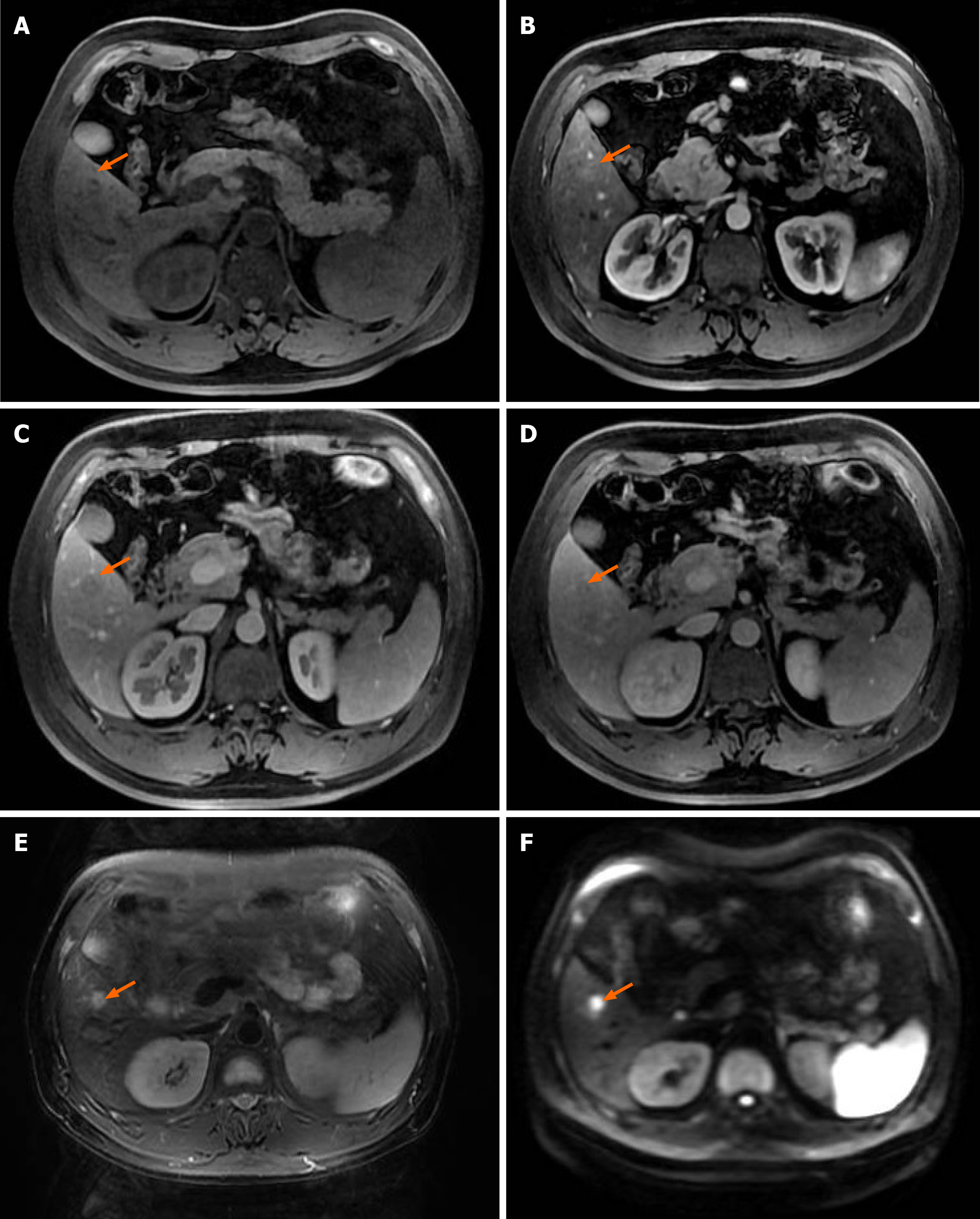
Figure 2 Magnetic resonance imaging of upper abdomen.
Magnetic resonance imaging scan revealed an iso T1 and long T2 signals, lesion only showed slightly enhancement in arterial phase, relatively low signal in portal and delayed phase hyperintense signal was observed in diffusion-weighted imaging. A: Iso T1 signal; B: Lesion only showed slightly enhancement in arterial phase; C and D: Relatively low signal in portal and delayed phase; E: Long T2 signal; F: Hyperintense signal was observed in diffusion-weighted imaging.
- Citation: Wang ZD, Haitham S, Gong JP, Pen ZL. Contrast enhanced ultrasound in diagnosing liver lesion that spontaneously disappeared: A case report . World J Clin Cases 2021; 9(21): 5948-5954
- URL: https://www.wjgnet.com/2307-8960/full/v9/i21/5948.htm
- DOI: https://dx.doi.org/10.12998/wjcc.v9.i21.5948


