Copyright
©The Author(s) 2021.
World J Clin Cases. Jul 26, 2021; 9(21): 5830-5839
Published online Jul 26, 2021. doi: 10.12998/wjcc.v9.i21.5830
Published online Jul 26, 2021. doi: 10.12998/wjcc.v9.i21.5830
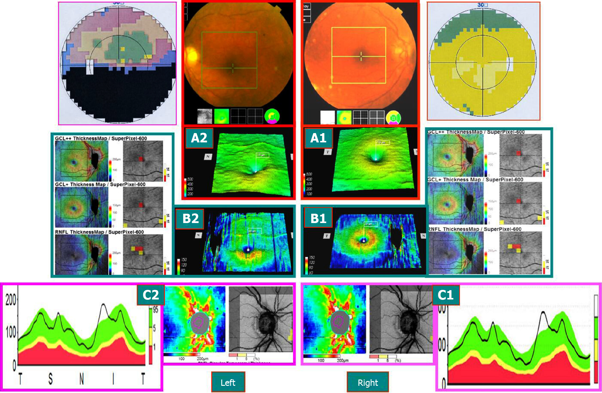
Figure 4 Sjögren’s syndrome patient on September 3, 2014 (typical case 2).
Hormones and immunosuppressants were administered to control the stability of the condition, and both eyes were subnormal by 3D-optical coherence tomography examination on December 31, 2013. A-C: Preclinical or latent subnormal eye of the right eye. The mGCC (A1 and B1) and the thickness of the nerve fibers around the optic disc (C1) were swollen and thickened, and no clinical symptoms were found. Late atrophy of the left eye AION disease (2 mo after onset): The mGCC delineated by horizontal sutures above the macular area were shrunk (A2 and B2), and optic disc showed a superior temporal nerve fiber atrophy (C2), which was consistent with that of the horizontal defect below the visual field. However, the mGCC under the macula of the left eye and thickness of the nerve fibers beneath the optic disc were still swollen and thickened (A2-C2). However, some nerve tract defects were noted above the field of vision, suggesting that the time may not be stable and might alter with the treatment.
- Citation: Zhang W, Sun XQ, Peng XY. Macular ganglion cell complex injury in different stages of anterior ischemic optic neuropathy. World J Clin Cases 2021; 9(21): 5830-5839
- URL: https://www.wjgnet.com/2307-8960/full/v9/i21/5830.htm
- DOI: https://dx.doi.org/10.12998/wjcc.v9.i21.5830


