Copyright
©The Author(s) 2021.
World J Clin Cases. Jul 26, 2021; 9(21): 5830-5839
Published online Jul 26, 2021. doi: 10.12998/wjcc.v9.i21.5830
Published online Jul 26, 2021. doi: 10.12998/wjcc.v9.i21.5830
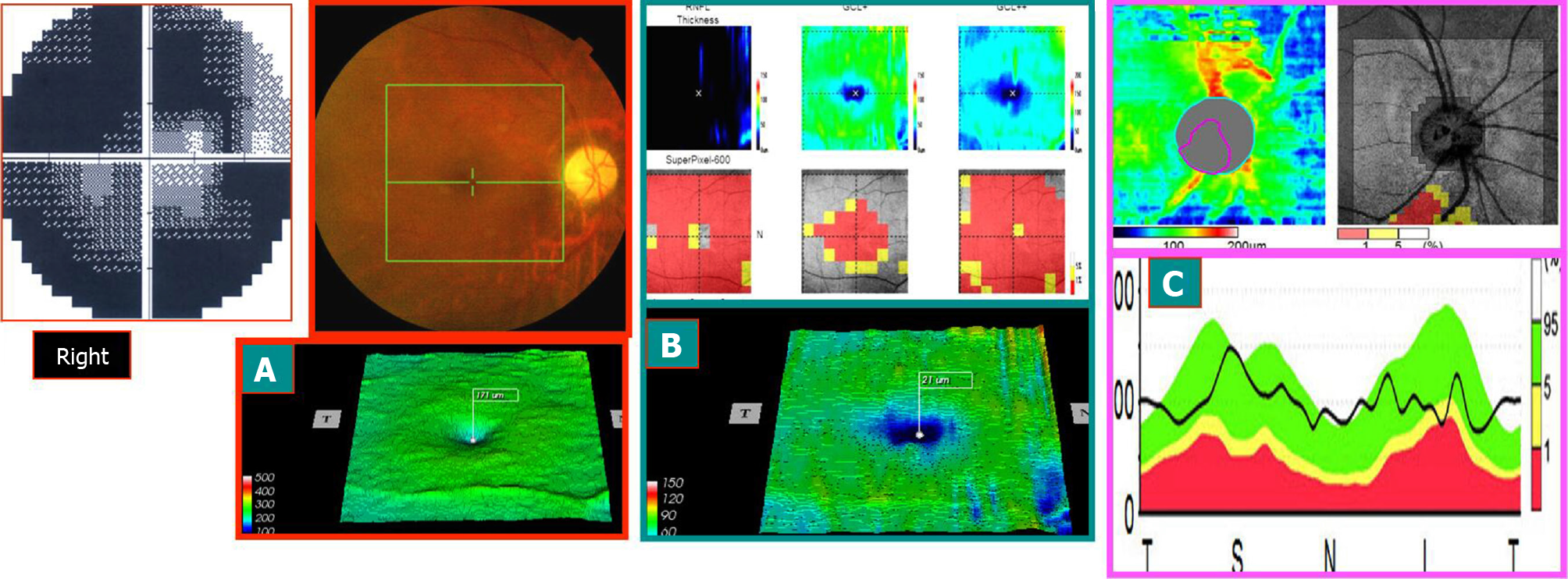
Figure 3 August 11, 2014, the stable atrophy of right eye in the late stage of onset (3 mo after onset): visual acuity was 0.
5 (typical case 1). A and B: Compared to Figure 2A and 2B, more shrinking and thinning were observed; C: Compared to Figure 2C, the nerve fibers around the optic disc were shrunk and thinned, especially in the lower part. This was consistent with the visual field showing that the horizontal defect above was more severe than the horizontal defect at the bottom. Right eye visual field: compared to that of the early stage of onset, the central area expanded. Hence, the visual acuity increased. The visual field of the left eye did not change before and after treatment.
- Citation: Zhang W, Sun XQ, Peng XY. Macular ganglion cell complex injury in different stages of anterior ischemic optic neuropathy. World J Clin Cases 2021; 9(21): 5830-5839
- URL: https://www.wjgnet.com/2307-8960/full/v9/i21/5830.htm
- DOI: https://dx.doi.org/10.12998/wjcc.v9.i21.5830


