Copyright
©The Author(s) 2021.
World J Clin Cases. Jul 26, 2021; 9(21): 5830-5839
Published online Jul 26, 2021. doi: 10.12998/wjcc.v9.i21.5830
Published online Jul 26, 2021. doi: 10.12998/wjcc.v9.i21.5830
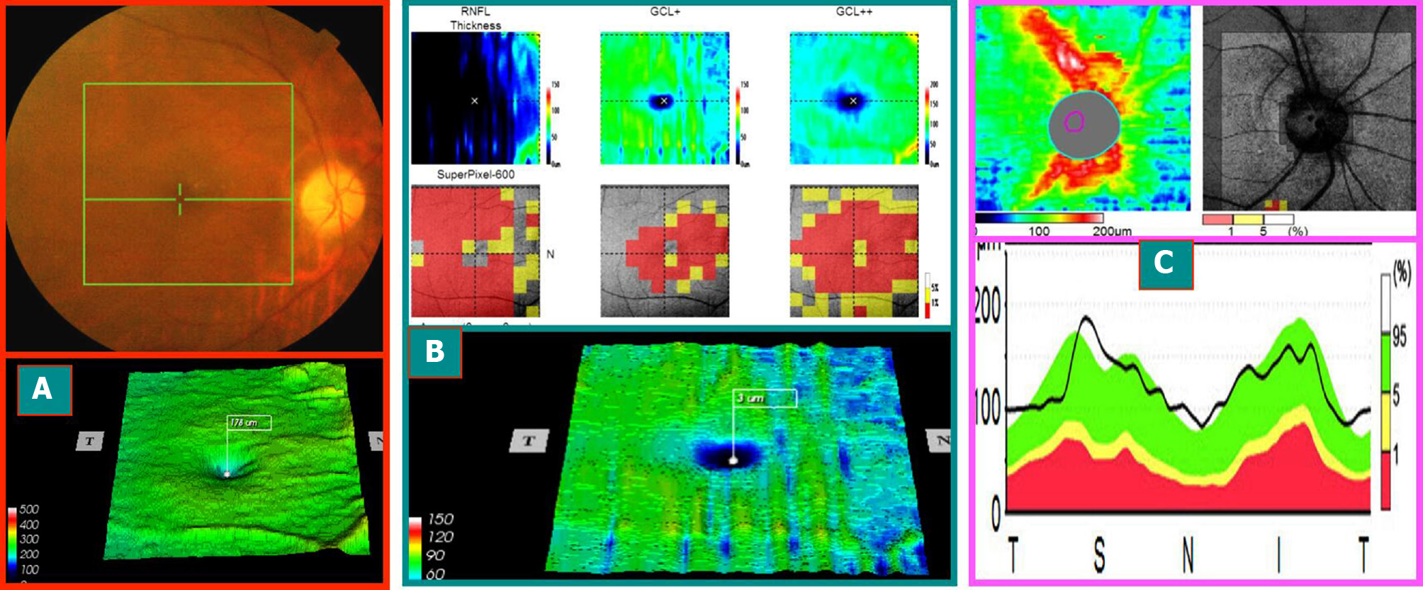
Figure 2 The mid-stage progression of the right eye and segregation phase (optical coherence tomography image at 1 mo after the onset of this case) (On June 9, 2014, typical case 1).
A: The optical disk was lighter, the boundary was distinct, and the circular area around the fovea of the macula had disappeared (obviously different from that of Figure 1A1); B: Macular ganglion cell complex ring disappeared, and three probabilistic maps (retinal nerve fiber layer, retinal ganglion cells + inner plexus layer, retinal nerve fiber layer + retinal ganglion cells + inner plexus layer) showed central red damage, which coincided with that of (A); C: The nerve fibers around the optic disc were swollen and thickened, and a few were beyond the normal high limit. The macular ganglion cell complex shrank and thinned. However, the nerve fibers around the optic disc were still swollen (swelling of the axons of the superficial ganglion cells), which occurred only 3 wk after onset and is known as the stage of progression-separation phenomenon in the middle stage of the onset. Moreover, the patient’s vision and visual field improved with treatment during this period.
- Citation: Zhang W, Sun XQ, Peng XY. Macular ganglion cell complex injury in different stages of anterior ischemic optic neuropathy. World J Clin Cases 2021; 9(21): 5830-5839
- URL: https://www.wjgnet.com/2307-8960/full/v9/i21/5830.htm
- DOI: https://dx.doi.org/10.12998/wjcc.v9.i21.5830


