Copyright
©The Author(s) 2021.
World J Clin Cases. Jul 16, 2021; 9(20): 5737-5743
Published online Jul 16, 2021. doi: 10.12998/wjcc.v9.i20.5737
Published online Jul 16, 2021. doi: 10.12998/wjcc.v9.i20.5737
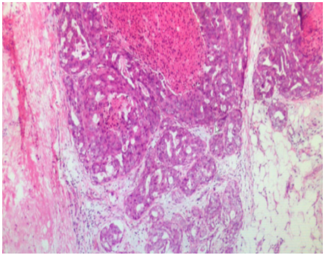
Figure 2 Postoperative pathological results: The tumor cells showed alveolar, solid, trabecular and other growth modes, and the tumor cells showed clear nucleoli and atypia.
Some areas of mitotic phase > 20/50 high-power field showed focal necrosis and partial mucinous degeneration. Tumor thrombus could be seen in the vessel. The mass size was 20 cm × 10 cm × 7 cm and was consistent with adrenocortical carcinoma. No renal invasion was observed. Tumor thrombus was observed in the renal vein. Immunohistochemical results revealed the following: S100(-), HMB-45(-), Melan-A (weakly positive), Inhibin-α (weakly positive), CgA(-), CKpan (local focus+), Vimentin(+), Syn(+), TFE3 (nucleus individual+), PAX-8(-), P53 (20% nucleus+), CD34(+), and Ki-67 (15%).
- Citation: Zhou Z, Luo HM, Tang J, Xu WJ, Wang BH, Peng XH, Tan H, Liu L, Long XY, Hong YD, Wu XB, Wang JP, Wang BQ, Xie HH, Fang Y, Luo Y, Li R, Wang Y. Multidisciplinary team therapy for left giant adrenocortical carcinoma: A case report. World J Clin Cases 2021; 9(20): 5737-5743
- URL: https://www.wjgnet.com/2307-8960/full/v9/i20/5737.htm
- DOI: https://dx.doi.org/10.12998/wjcc.v9.i20.5737


