Copyright
©The Author(s) 2021.
World J Clin Cases. Jul 16, 2021; 9(20): 5709-5716
Published online Jul 16, 2021. doi: 10.12998/wjcc.v9.i20.5709
Published online Jul 16, 2021. doi: 10.12998/wjcc.v9.i20.5709
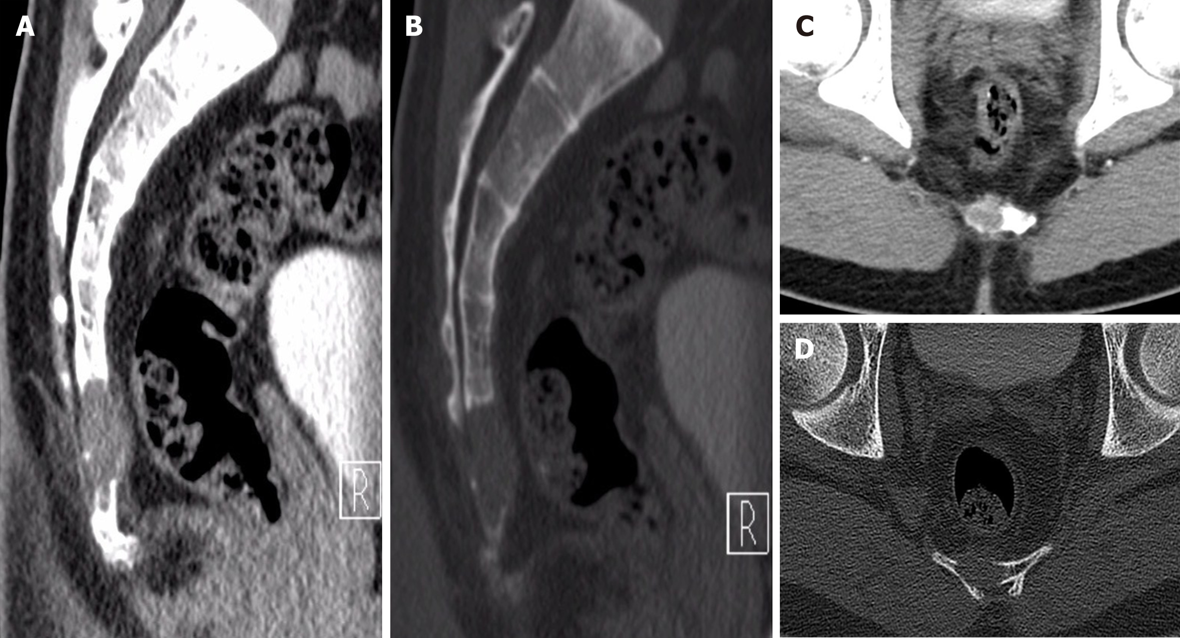
Figure 1 Computed tomography showed shows an osteolytic lesion with irregular margins and cortical breach.
A: First preoperative sagittal computed tomography (CT) images (bone window); B: First preoperative sagittal CT images (soft-tissue window); C: First preoperative axial CT images (bone window); D: First preoperative axial CT images (soft-tissue window).
- Citation: Zheng BW, Niu HQ, Wang XB, Li J. Sacral chondroblastoma — a rare location, a rare pathology: A case report and review of literature. World J Clin Cases 2021; 9(20): 5709-5716
- URL: https://www.wjgnet.com/2307-8960/full/v9/i20/5709.htm
- DOI: https://dx.doi.org/10.12998/wjcc.v9.i20.5709


