Copyright
©The Author(s) 2021.
World J Clin Cases. Jul 16, 2021; 9(20): 5668-5674
Published online Jul 16, 2021. doi: 10.12998/wjcc.v9.i20.5668
Published online Jul 16, 2021. doi: 10.12998/wjcc.v9.i20.5668
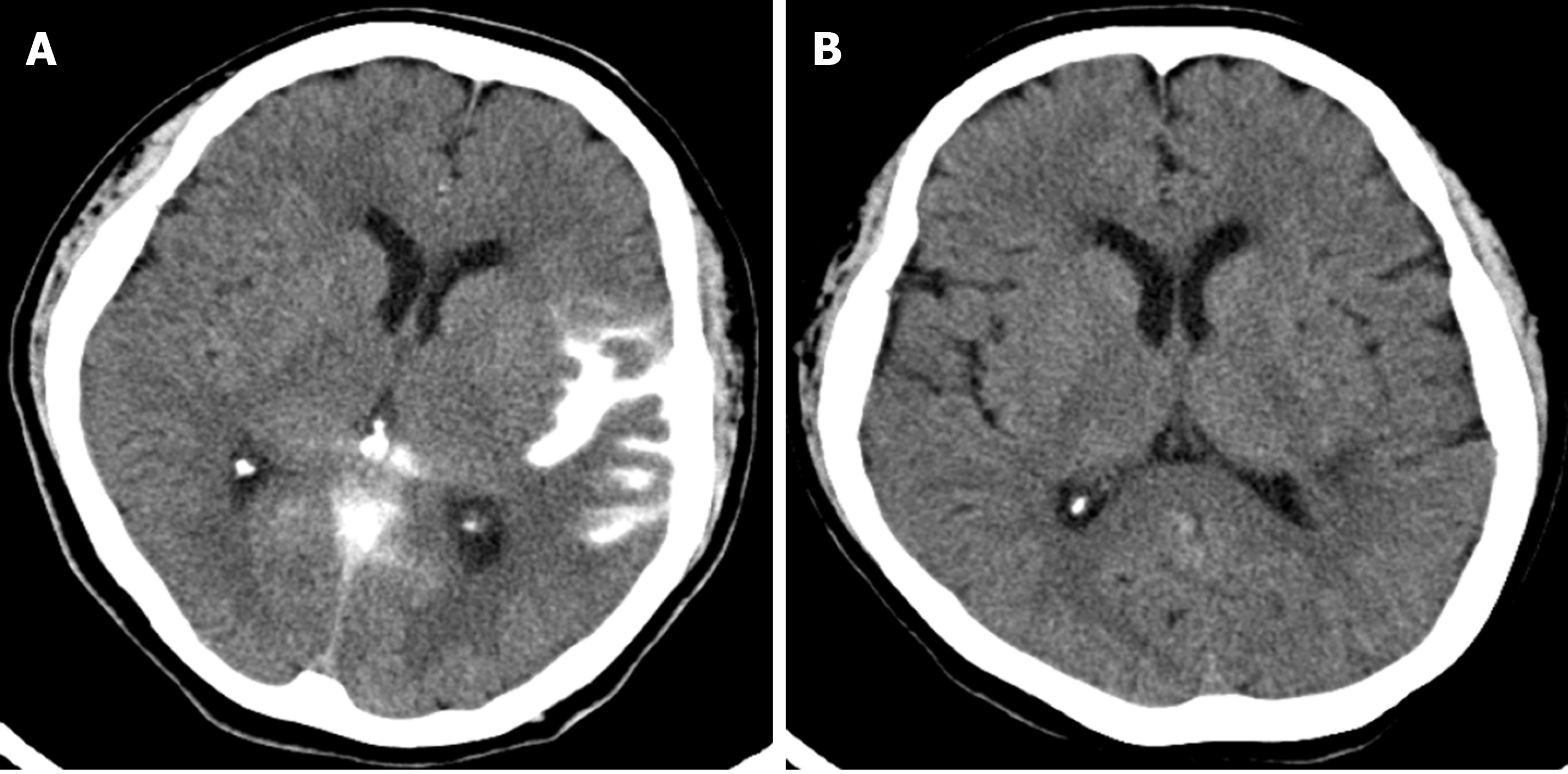
Figure 3 Immediate post-procedure and 12-d follow-up brain computed tomography images.
A: Immediately after the procedure, subarachnoid hemorrhage was identified in the left sylvian cistern on brain computed tomography (CT); B: On the follow-up CT 12 d after the procedure, the subarachnoid hemorrhage was resolved, and a focal low density of infarction was seen in the temporal lobe.
- Citation: Kang JY, Yi KS, Cha SH, Choi CH, Kim Y, Lee J, Cho BS. Gelfoam embolization for distal, medium vessel injury during mechanical thrombectomy in acute stroke: A case report. World J Clin Cases 2021; 9(20): 5668-5674
- URL: https://www.wjgnet.com/2307-8960/full/v9/i20/5668.htm
- DOI: https://dx.doi.org/10.12998/wjcc.v9.i20.5668


