Copyright
©The Author(s) 2021.
World J Clin Cases. Jul 16, 2021; 9(20): 5668-5674
Published online Jul 16, 2021. doi: 10.12998/wjcc.v9.i20.5668
Published online Jul 16, 2021. doi: 10.12998/wjcc.v9.i20.5668
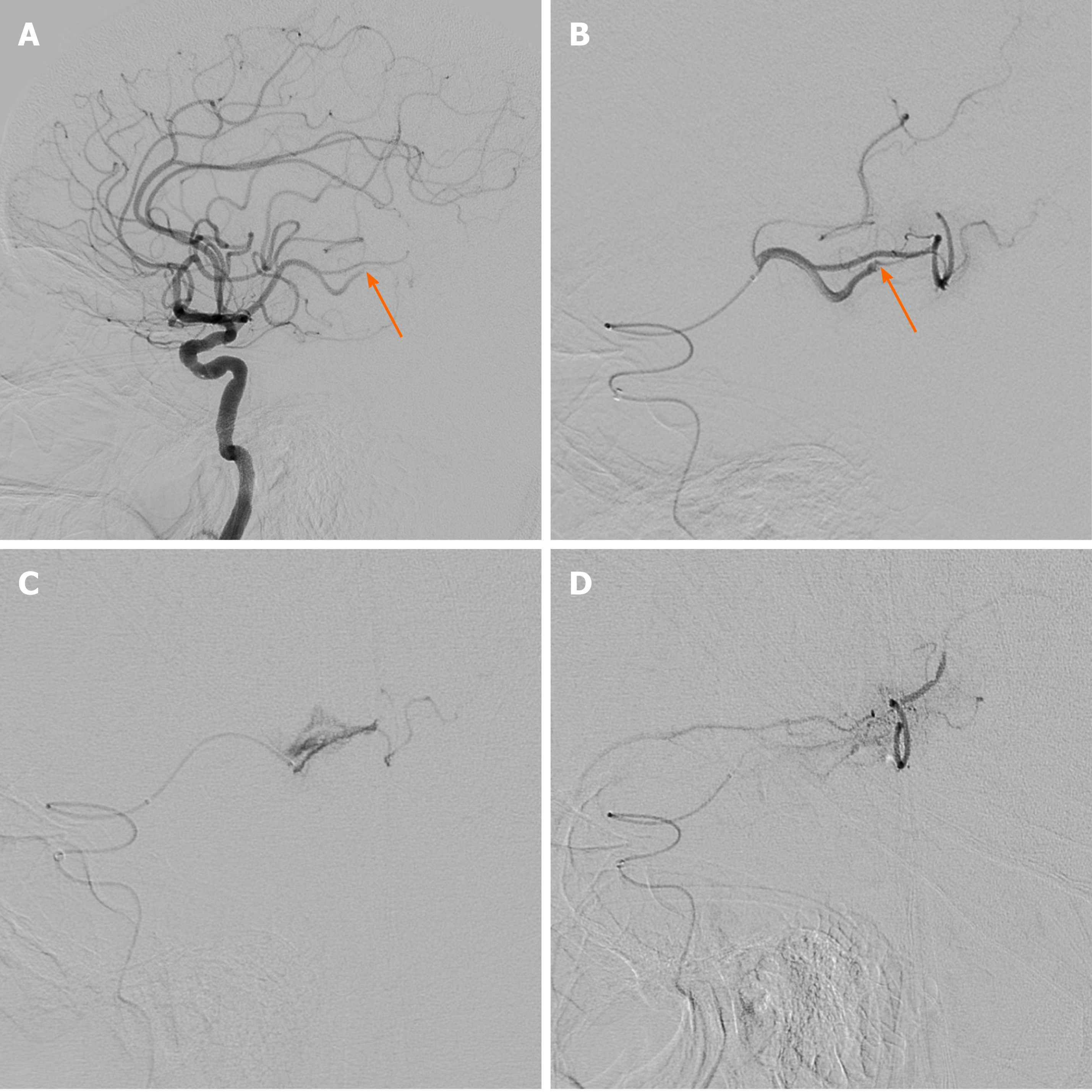
Figure 2 Cerebral angiography and endovascular management.
A and B: Initial cerebral angiography revealed occlusion of the inferior division of the middle cerebral artery at the M2-3 segment (arrow); C: During advancement of the microcatheter, resistance was encountered at the occlusion point, and the tension of the microcatheter was released. Cautious angiography was performed, revealing extravasation at the M3 branch due to vascular injury; D: Extravasation of contrast medium persisted in delayed angiography, and embolization was performed using gelfoam (1400-2000 μm). Repeat angiography confirmed hemostasis of the injured vessel.
- Citation: Kang JY, Yi KS, Cha SH, Choi CH, Kim Y, Lee J, Cho BS. Gelfoam embolization for distal, medium vessel injury during mechanical thrombectomy in acute stroke: A case report. World J Clin Cases 2021; 9(20): 5668-5674
- URL: https://www.wjgnet.com/2307-8960/full/v9/i20/5668.htm
- DOI: https://dx.doi.org/10.12998/wjcc.v9.i20.5668


