Copyright
©The Author(s) 2021.
World J Clin Cases. Jul 16, 2021; 9(20): 5637-5646
Published online Jul 16, 2021. doi: 10.12998/wjcc.v9.i20.5637
Published online Jul 16, 2021. doi: 10.12998/wjcc.v9.i20.5637
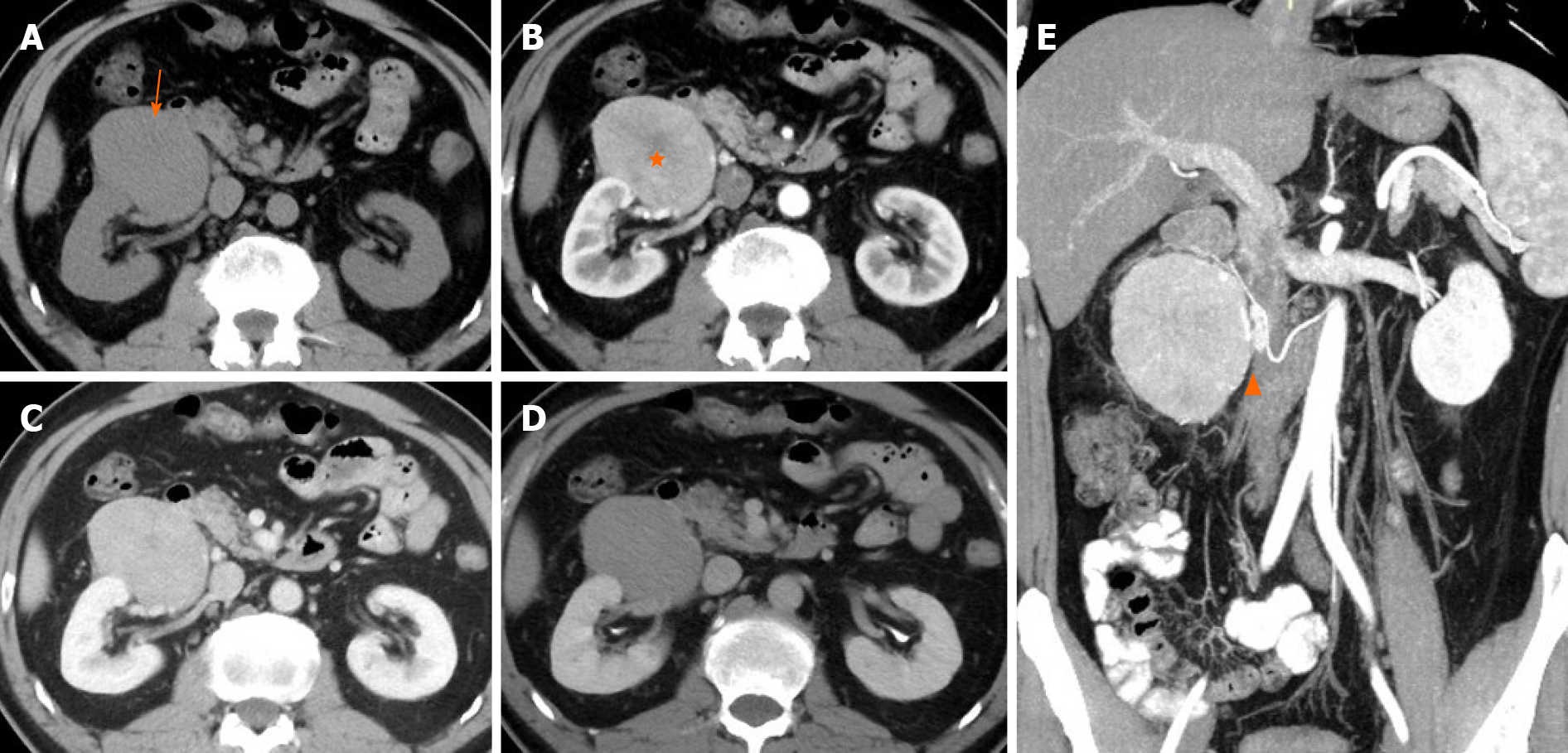
Figure 1 Computed tomography.
A: Pre-contrast image in the transverse plane showed that the density of mass was 40 HU (arrow); B-D: On a contrast-enhanced scan, the tumor presented uneven high enhancement with a stellate central scar in arterial phase (B, 111 HU, orange star) and withdrawal enhancement uniformly in portal vein (C, 95 HU) and delayed phases (D, 79 HU); E: On multiplanar reconstruction, the blood supply of the tumor derives from the right testicular artery (orange arrowhead).
- Citation: Wei YY, Li Y, Shi YJ, Li XT, Sun YS. Primary extra-pancreatic pancreatic-type acinar cell carcinoma in the right perinephric space: A case report and review of literature. World J Clin Cases 2021; 9(20): 5637-5646
- URL: https://www.wjgnet.com/2307-8960/full/v9/i20/5637.htm
- DOI: https://dx.doi.org/10.12998/wjcc.v9.i20.5637


