Copyright
©The Author(s) 2021.
World J Clin Cases. Jul 16, 2021; 9(20): 5594-5604
Published online Jul 16, 2021. doi: 10.12998/wjcc.v9.i20.5594
Published online Jul 16, 2021. doi: 10.12998/wjcc.v9.i20.5594
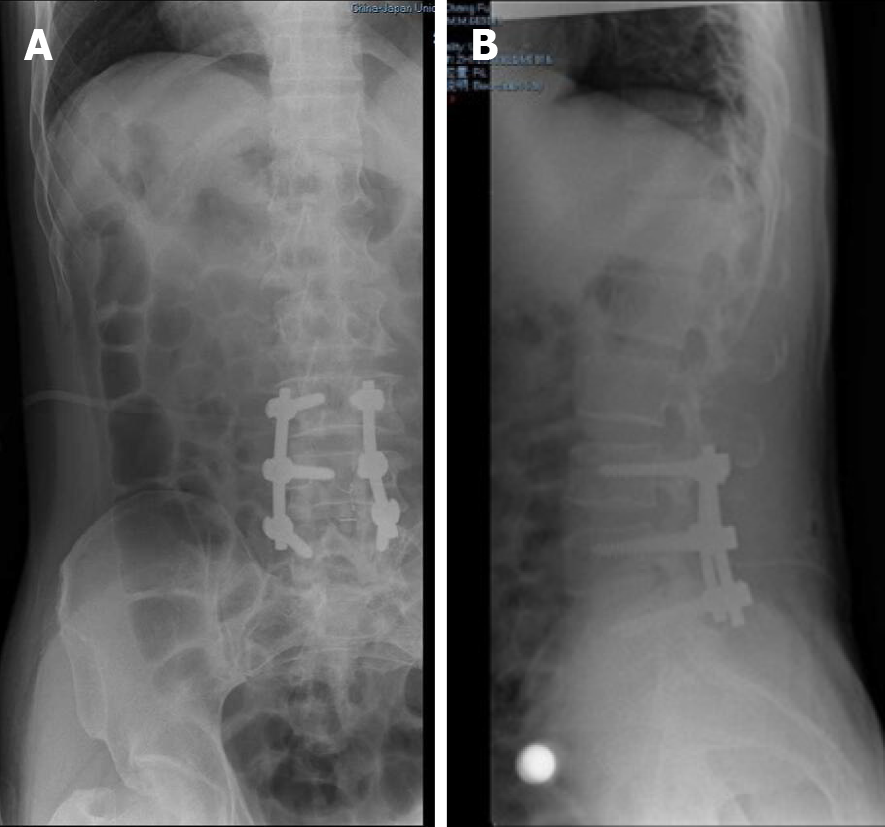
Figure 5 Postoperative X-ray at the patient’s first admission.
A and B: Anteroposterior (A) and lateral (B) radiographs showed that the internal fixative was well positioned in the patient.
- Citation: Ouyang Y, Qu Y, Dong RP, Kang MY, Yu T, Cheng XL, Zhao JW. Spinal dural arteriovenous fistula 8 years after lumbar discectomy surgery: A case report and review of literature. World J Clin Cases 2021; 9(20): 5594-5604
- URL: https://www.wjgnet.com/2307-8960/full/v9/i20/5594.htm
- DOI: https://dx.doi.org/10.12998/wjcc.v9.i20.5594


