Copyright
©The Author(s) 2021.
World J Clin Cases. Jul 16, 2021; 9(20): 5594-5604
Published online Jul 16, 2021. doi: 10.12998/wjcc.v9.i20.5594
Published online Jul 16, 2021. doi: 10.12998/wjcc.v9.i20.5594
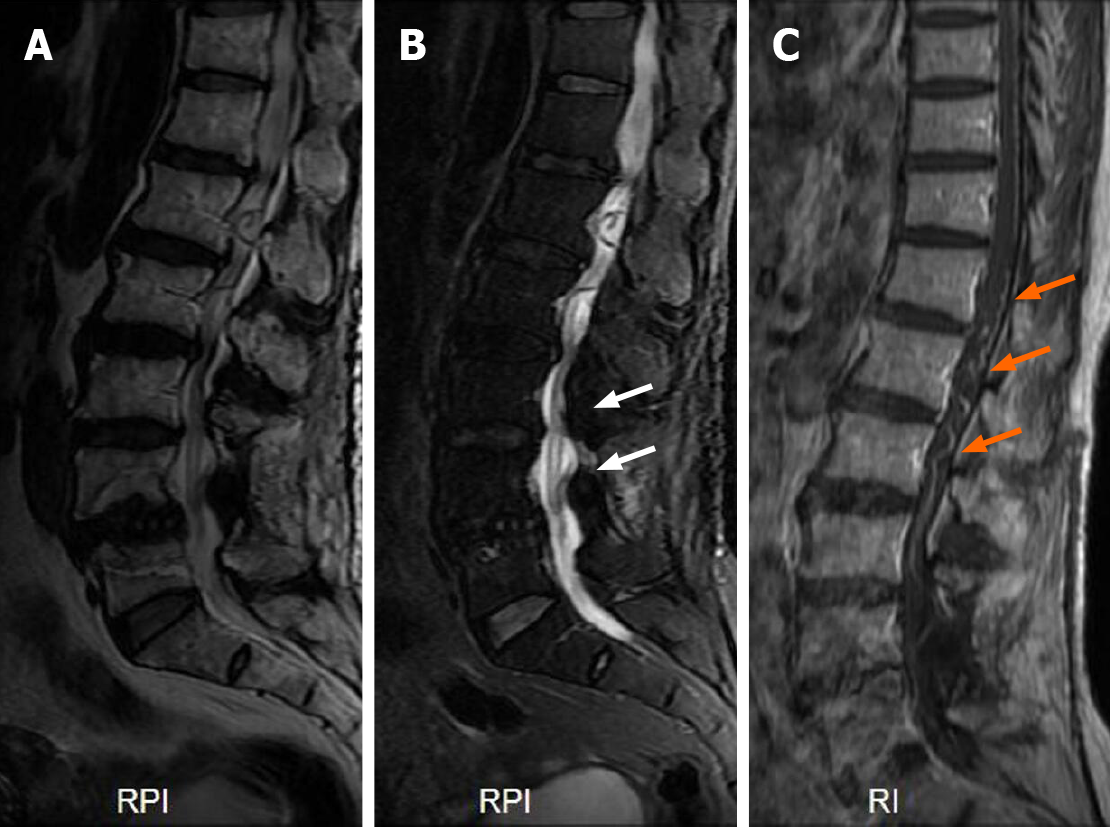
Figure 3 Preoperative magnetic resonance imaging at second admission.
A and B: On T2 image, L4 vertebral body was displaced forward, the edge of vertebral body was irregular, multi segment intervertebral disc protruded backward (A), and high signal shadow was seen in the spinal cord (white arrows) (B); C: Enhanced magnetic resonance imaging showed tortuous dilated vessels in the dorsal side of the spinal cord (orange arrows). RPI: Right posterior image; RI: Right image.
- Citation: Ouyang Y, Qu Y, Dong RP, Kang MY, Yu T, Cheng XL, Zhao JW. Spinal dural arteriovenous fistula 8 years after lumbar discectomy surgery: A case report and review of literature. World J Clin Cases 2021; 9(20): 5594-5604
- URL: https://www.wjgnet.com/2307-8960/full/v9/i20/5594.htm
- DOI: https://dx.doi.org/10.12998/wjcc.v9.i20.5594


