Copyright
©The Author(s) 2021.
World J Clin Cases. Jul 16, 2021; 9(20): 5594-5604
Published online Jul 16, 2021. doi: 10.12998/wjcc.v9.i20.5594
Published online Jul 16, 2021. doi: 10.12998/wjcc.v9.i20.5594
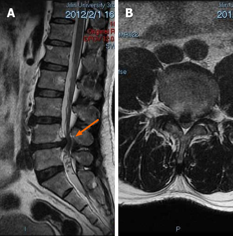
Figure 1 Patient’s first admission preoperative magnetic resonance imaging.
A: Sagittal view of the patient showed a straightening of the physiological curvature of the lumbar spine and a herniated disc at L4-L5 segments (orange arrow); B: Axial scan showed a herniated disc at segments L4-L5, compression of the dura, marked compression of the foramina on both sides and spinal canal stenosis.
- Citation: Ouyang Y, Qu Y, Dong RP, Kang MY, Yu T, Cheng XL, Zhao JW. Spinal dural arteriovenous fistula 8 years after lumbar discectomy surgery: A case report and review of literature. World J Clin Cases 2021; 9(20): 5594-5604
- URL: https://www.wjgnet.com/2307-8960/full/v9/i20/5594.htm
- DOI: https://dx.doi.org/10.12998/wjcc.v9.i20.5594


