Copyright
©The Author(s) 2021.
World J Clin Cases. Jan 16, 2021; 9(2): 509-515
Published online Jan 16, 2021. doi: 10.12998/wjcc.v9.i2.509
Published online Jan 16, 2021. doi: 10.12998/wjcc.v9.i2.509
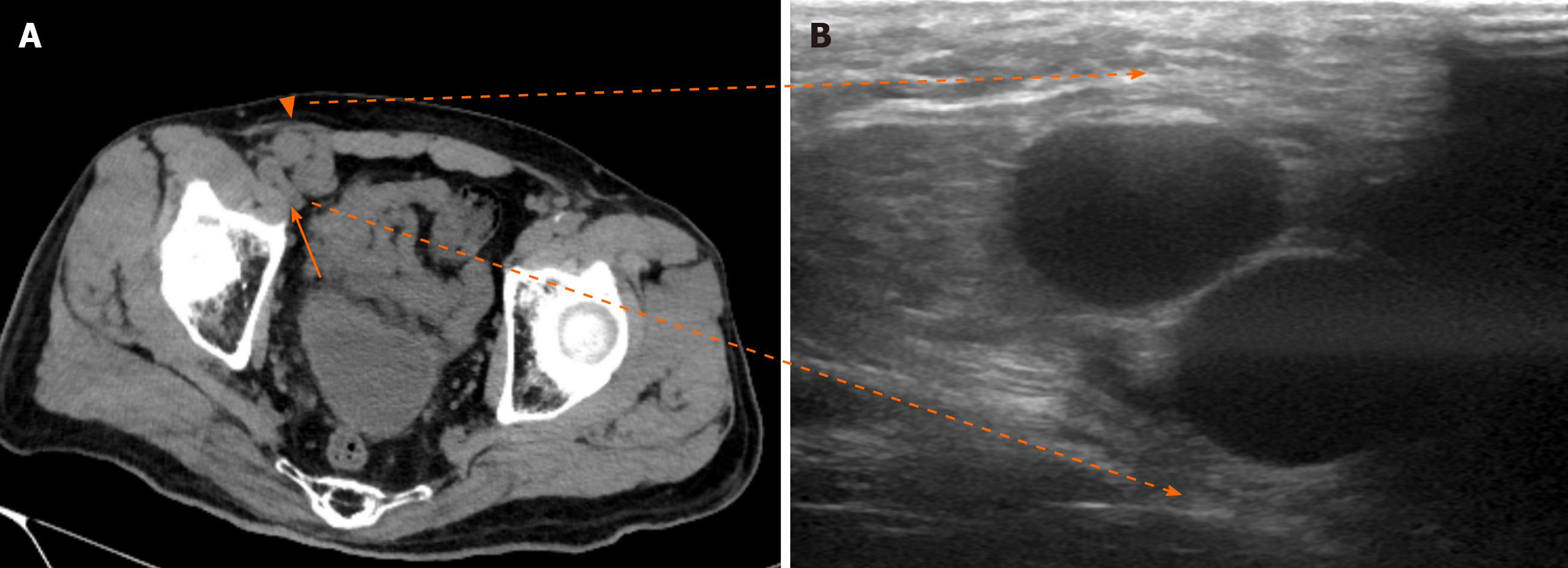
Figure 2 Preoperative computed tomography and ultrasound findings.
A: Preoperative computed tomography revealed a portosystemic venous shunt vessel located in the ventral part of the femoral vein (arrow) and entered the internal inguinal ring (triangle); B: Abdominal ultrasound detected the shunt vessel just under the groin.
- Citation: Yura M, Yo K, Hara A, Hayashi K, Tajima Y, Kaneko Y, Fujisaki H, Hirata A, Takano K, Hongo K, Yoneyama K, Nakagawa M. Indirect inguinal hernia containing portosystemic shunt vessel: A case report. World J Clin Cases 2021; 9(2): 509-515
- URL: https://www.wjgnet.com/2307-8960/full/v9/i2/509.htm
- DOI: https://dx.doi.org/10.12998/wjcc.v9.i2.509


