Copyright
©The Author(s) 2021.
World J Clin Cases. Jan 16, 2021; 9(2): 489-495
Published online Jan 16, 2021. doi: 10.12998/wjcc.v9.i2.489
Published online Jan 16, 2021. doi: 10.12998/wjcc.v9.i2.489
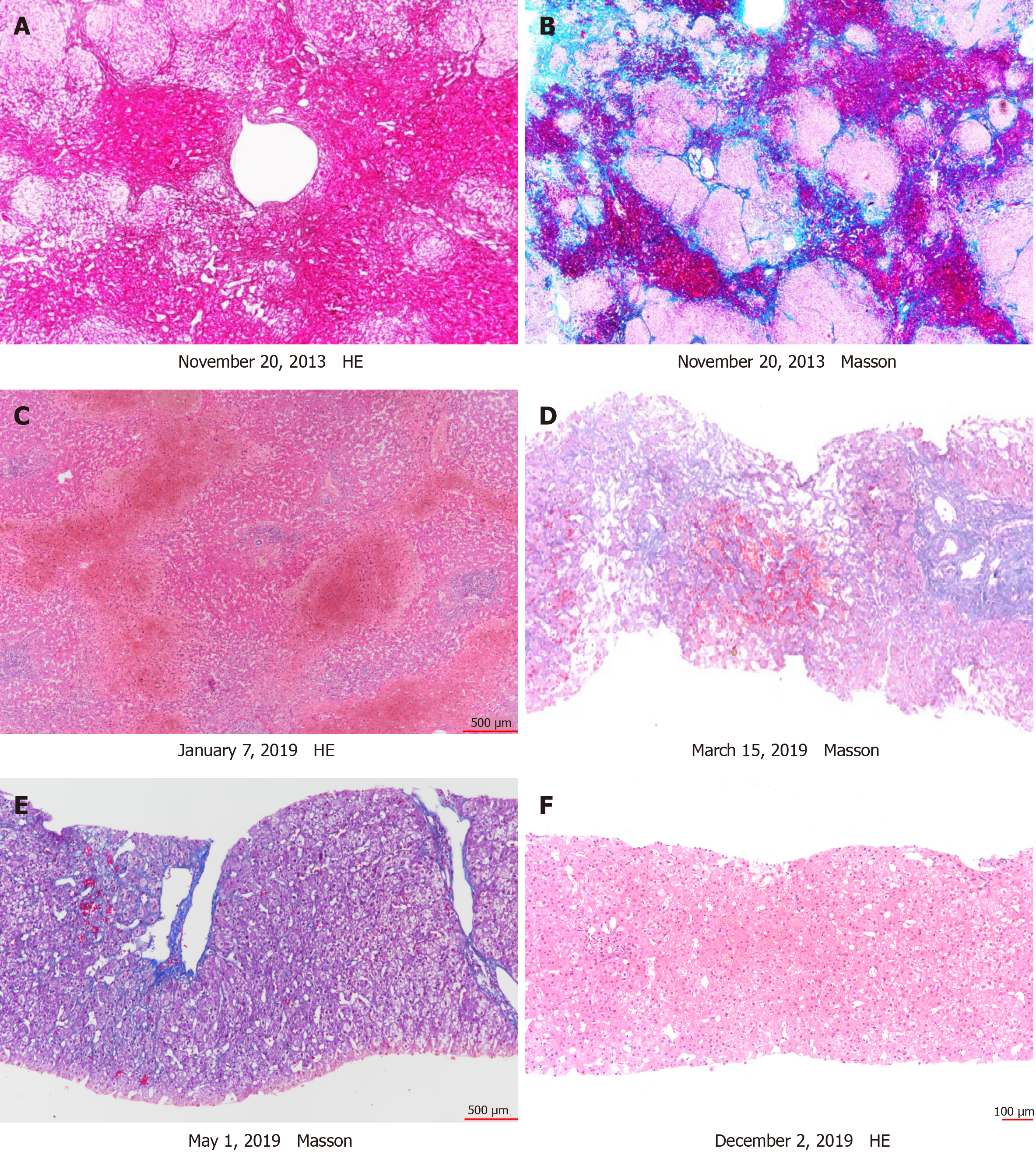
Figure 1 Hepatic venography.
A and B: Native liver showed marked sinusoidal dilatation and congestion in centrilobular regions and extensive bridging fibrosis and necrosis linking central to central areas; C: Explanted first liver graft characterized by massive perivenular congestion and hemorrhage with marked sinusoidal dilatation. Portal tract was not remarkable; D: Two months after liver retransplantation, liver biopsy was performed to clarify the diagnosis. The second liver graft liver pathology showed sinusoidal dilatation and congestion; E: In addition to warfarin, tacrolimus was switched to cyclosporine A. Two months after treatment, perivenular congestion and sinusoidal dilation were alleviated and were only observed in the focal perivenular area; F: Nine months later, there was no perivenular congestion and only mild sinusoidal dilatation.
- Citation: Liu Y, Sun LY, Zhu ZJ, Wei L, Qu W, Zeng ZG. Is sinusoidal obstructive syndrome a recurrent disease after liver transplantation? A case report . World J Clin Cases 2021; 9(2): 489-495
- URL: https://www.wjgnet.com/2307-8960/full/v9/i2/489.htm
- DOI: https://dx.doi.org/10.12998/wjcc.v9.i2.489


