Copyright
©The Author(s) 2021.
World J Clin Cases. Jan 16, 2021; 9(2): 445-456
Published online Jan 16, 2021. doi: 10.12998/wjcc.v9.i2.445
Published online Jan 16, 2021. doi: 10.12998/wjcc.v9.i2.445
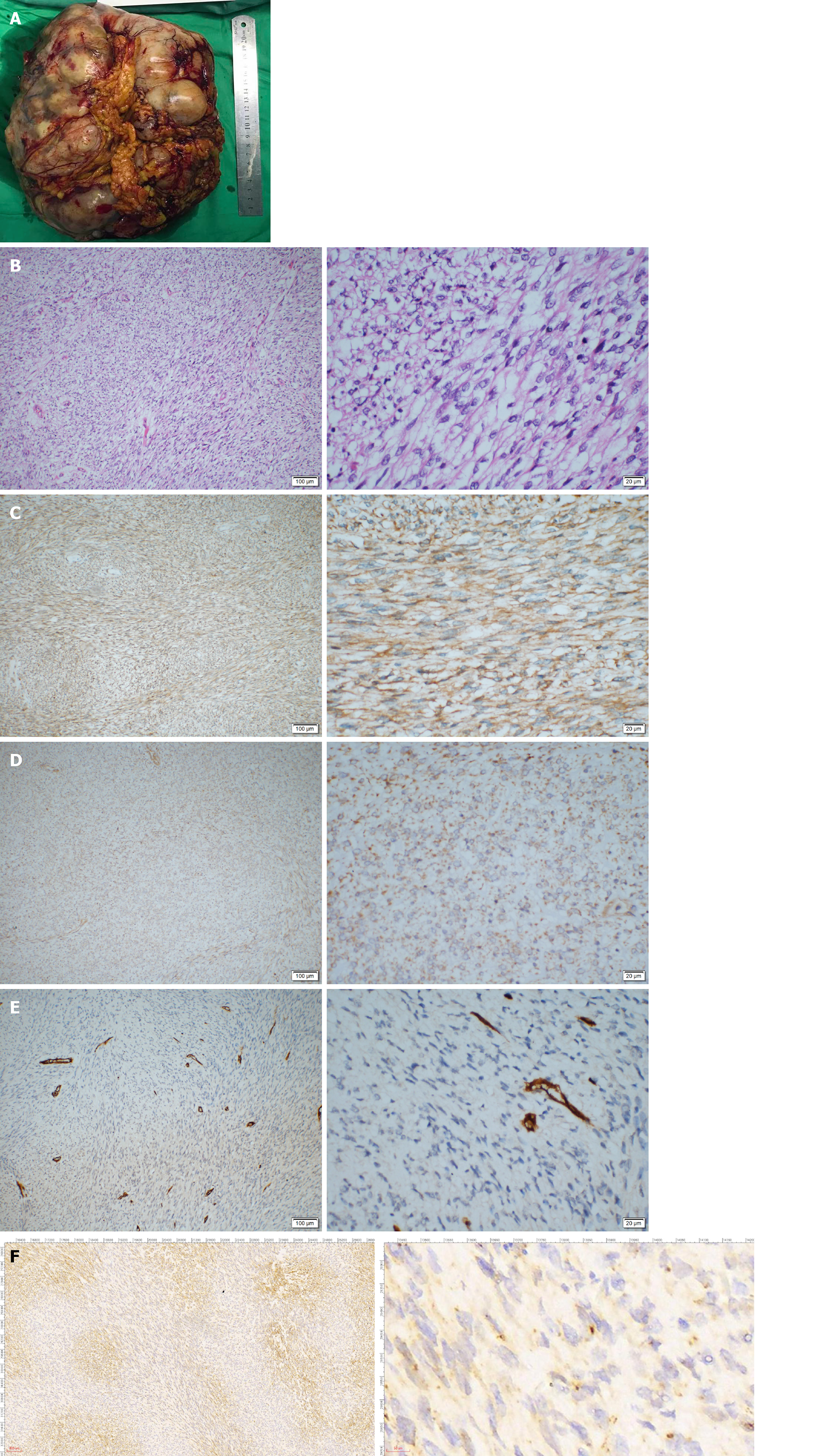
Figure 3 Pathological examination of the tumor.
A: The gross morphology of the resected tumor specimen was huge (27 cm × 21 cm × 9 cm) with necrosis and hemorrhage on the surface; B: Pathological examination showed hypercellularity with spindle cells, and a high mitotic activity with a rate of 30/10 high power field (HPF); C and D: On immunohistochemistry staining, BCL-2 and DOG-1 were weakly positive; E and F: CD34 and STAT6 were negative.
- Citation: Guo YC, Yao LY, Tian ZS, Shi B, Liu Y, Wang YY. Malignant solitary fibrous tumor of the greater omentum: A case report and review of literature. World J Clin Cases 2021; 9(2): 445-456
- URL: https://www.wjgnet.com/2307-8960/full/v9/i2/445.htm
- DOI: https://dx.doi.org/10.12998/wjcc.v9.i2.445


