Copyright
©The Author(s) 2021.
World J Clin Cases. Jan 16, 2021; 9(2): 445-456
Published online Jan 16, 2021. doi: 10.12998/wjcc.v9.i2.445
Published online Jan 16, 2021. doi: 10.12998/wjcc.v9.i2.445
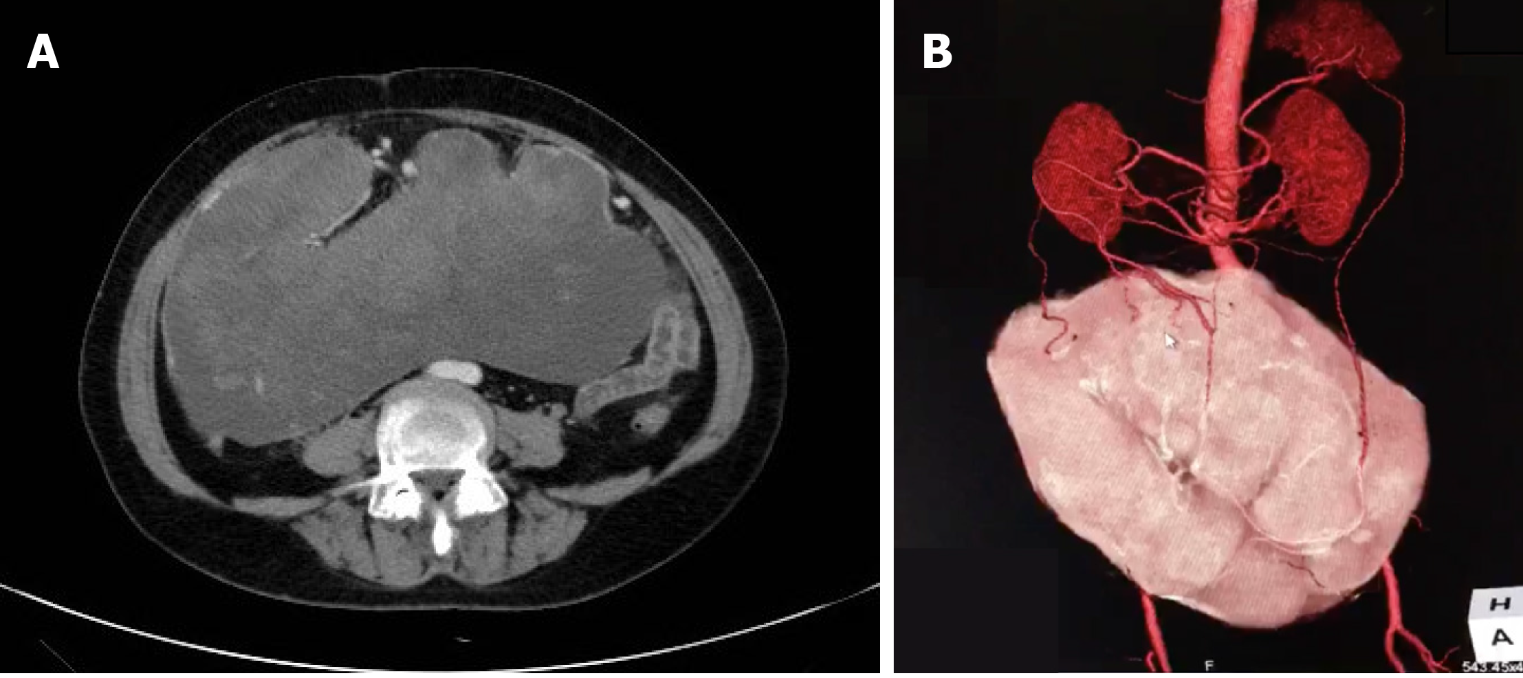
Figure 1 Imaging of the abdominal mass.
A: Contrast-enhanced abdominal computed tomography (CT) scan showing a huge mass measuring 25.4 cm × 23.0 cm with mixed density and heterogeneous enhancement (arrow); B: CT 3D reconstruction showing that the feeding arteries were from the splenic artery, right colic artery, and middle colic artery (arrows).
- Citation: Guo YC, Yao LY, Tian ZS, Shi B, Liu Y, Wang YY. Malignant solitary fibrous tumor of the greater omentum: A case report and review of literature. World J Clin Cases 2021; 9(2): 445-456
- URL: https://www.wjgnet.com/2307-8960/full/v9/i2/445.htm
- DOI: https://dx.doi.org/10.12998/wjcc.v9.i2.445


