Copyright
©The Author(s) 2021.
World J Clin Cases. Jul 6, 2021; 9(19): 5287-5293
Published online Jul 6, 2021. doi: 10.12998/wjcc.v9.i19.5287
Published online Jul 6, 2021. doi: 10.12998/wjcc.v9.i19.5287
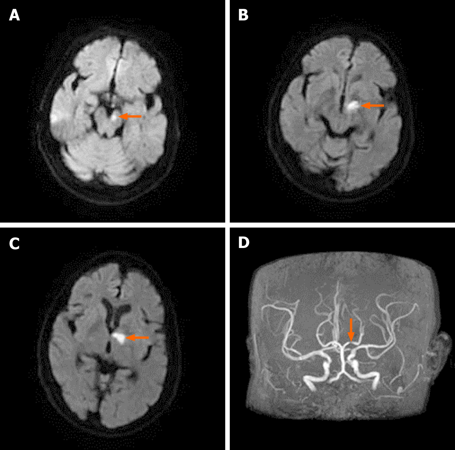
Figure 1 Magnetic resonance imaging and magnetic resonance angiography of the brain.
A-C: Diffusion-weighted imaging showing cerebral infarction (orange arrows). A: Infarction in the left cerebral peduncle zone; B: Infarction in the junctional region of the left dorsal thalamus and cerebral peduncle; C: Infarction in the left dorsal thalamus; D: Magnetic resonance angiography showing severe stenosis of the P1 segment of the left posterior cerebral artery (orange arrow).
- Citation: Li ZS, Fang JJ, Xiang XH, Zhao GH. Hemichorea due to ipsilateral thalamic infarction: A case report. World J Clin Cases 2021; 9(19): 5287-5293
- URL: https://www.wjgnet.com/2307-8960/full/v9/i19/5287.htm
- DOI: https://dx.doi.org/10.12998/wjcc.v9.i19.5287


