Copyright
©The Author(s) 2021.
World J Clin Cases. Jul 6, 2021; 9(19): 5259-5265
Published online Jul 6, 2021. doi: 10.12998/wjcc.v9.i19.5259
Published online Jul 6, 2021. doi: 10.12998/wjcc.v9.i19.5259
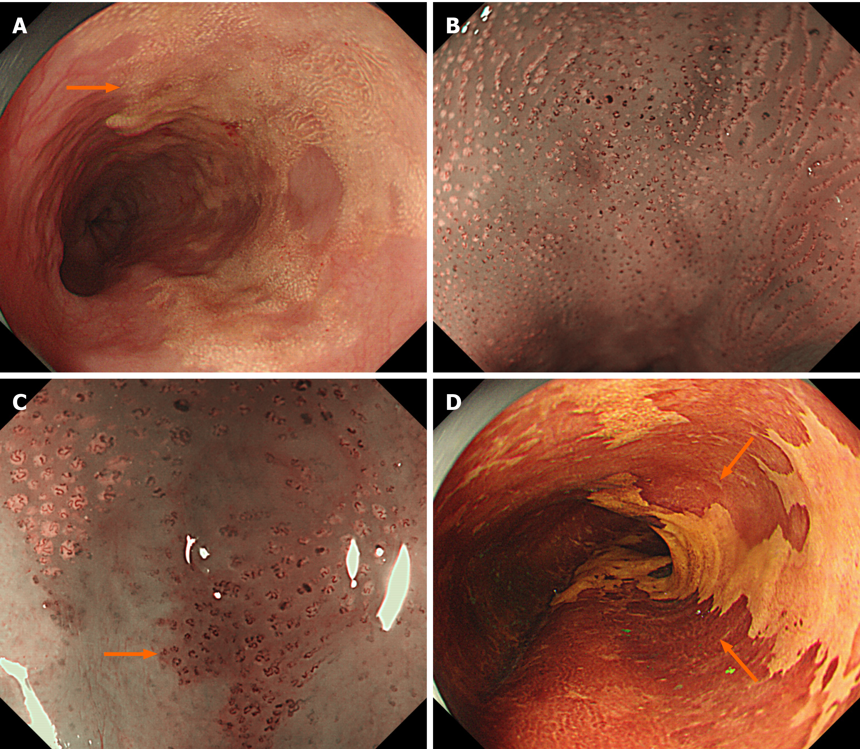
Figure 1 Endoscopic illustrations (GIF-H260Z, Olympus).
A: White light endoscopy showed a semi-circumferential, irregular, yellowish-colored, and granular lesion localized in the middle and lower esophagus (orange arrow); B: Narrow band imaging endoscopy revealed aggregation of minute yellowish spots with tortuous microvessels inside; C: Type B1 intrapapillary capillary loops were identified by magnifying endoscopy in the region around the yellow spots, and the lesion was positive for background coloration (orange arrow); D: Lugol's iodine staining revealed a well-demarcated unstained lesion (orange arrow).
- Citation: Yang XY, Fu KI, Chen YP, Chen ZW, Ding J. Diffuse xanthoma in early esophageal cancer: A case report. World J Clin Cases 2021; 9(19): 5259-5265
- URL: https://www.wjgnet.com/2307-8960/full/v9/i19/5259.htm
- DOI: https://dx.doi.org/10.12998/wjcc.v9.i19.5259


