Copyright
©The Author(s) 2021.
World J Clin Cases. Jul 6, 2021; 9(19): 5211-5216
Published online Jul 6, 2021. doi: 10.12998/wjcc.v9.i19.5211
Published online Jul 6, 2021. doi: 10.12998/wjcc.v9.i19.5211
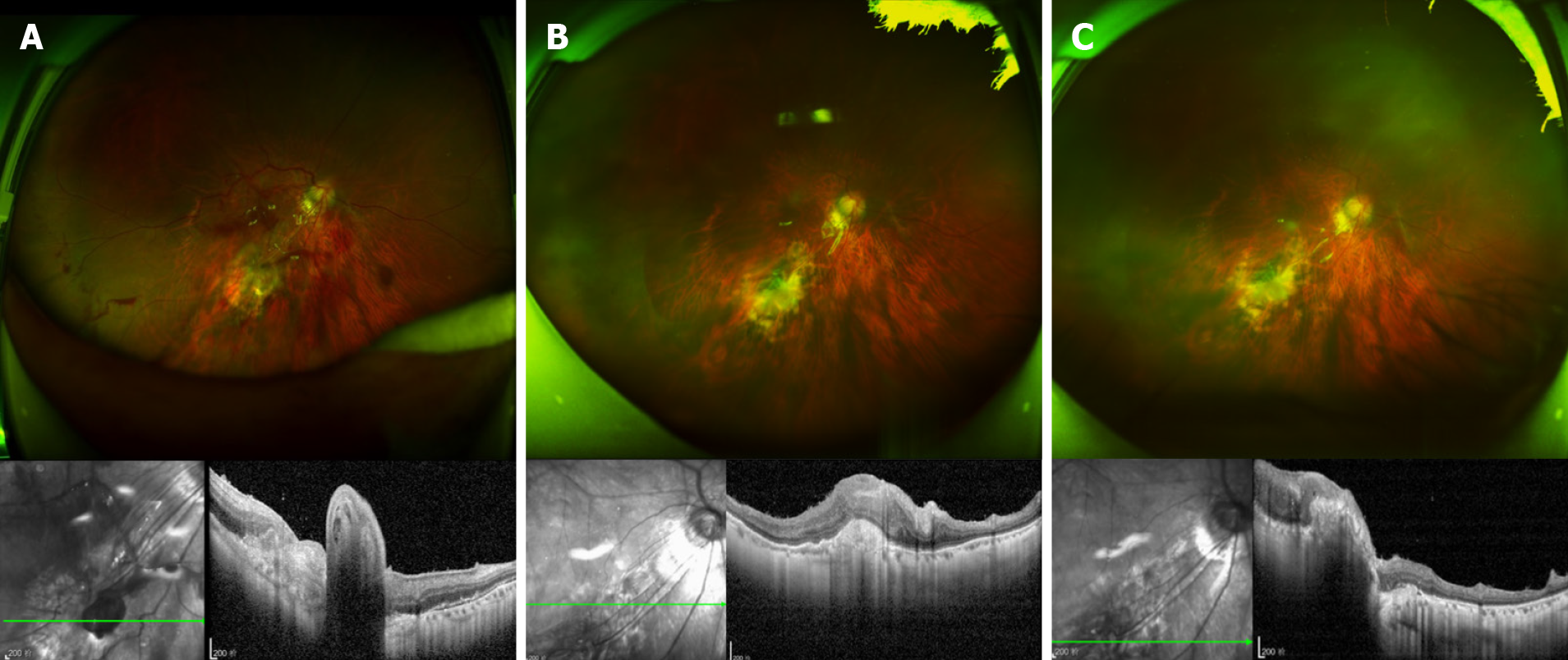
Figure 5 Ophthalmological examination after surgery A: Opel fundus image and optical coherence tomography (OCT) at 2 wk after surgery.
The Opel fundus image shows that the autophagic fascia was in place in the submacrotemporal laceration of the right eye, with the hole closed and the retina flat. OCT examination showed that the stuffing tissue was protruded in place at the laceration, and the surrounding retina was flattened; B and C: Opel fundus images and OCT at 5 mo after surgery and 2 mo after removal of the silicone oil. The Opel fundus image shows that the autophagic fascia was in place in the submacrotemporal laceration of the right eye, with the hole closed and the retina flat. OCT showed that the stuffing tissue in the laceration was in place and connected with the surrounding tissue, and the surrounding retina was flat.
- Citation: Yi QY, Wang SS, Gui Q, Chen LS, Li WD. Autologous tenon capsule packing to treat posterior exit wound of penetrating injury: A case report. World J Clin Cases 2021; 9(19): 5211-5216
- URL: https://www.wjgnet.com/2307-8960/full/v9/i19/5211.htm
- DOI: https://dx.doi.org/10.12998/wjcc.v9.i19.5211


