Copyright
©The Author(s) 2021.
World J Clin Cases. Jul 6, 2021; 9(19): 5197-5202
Published online Jul 6, 2021. doi: 10.12998/wjcc.v9.i19.5197
Published online Jul 6, 2021. doi: 10.12998/wjcc.v9.i19.5197
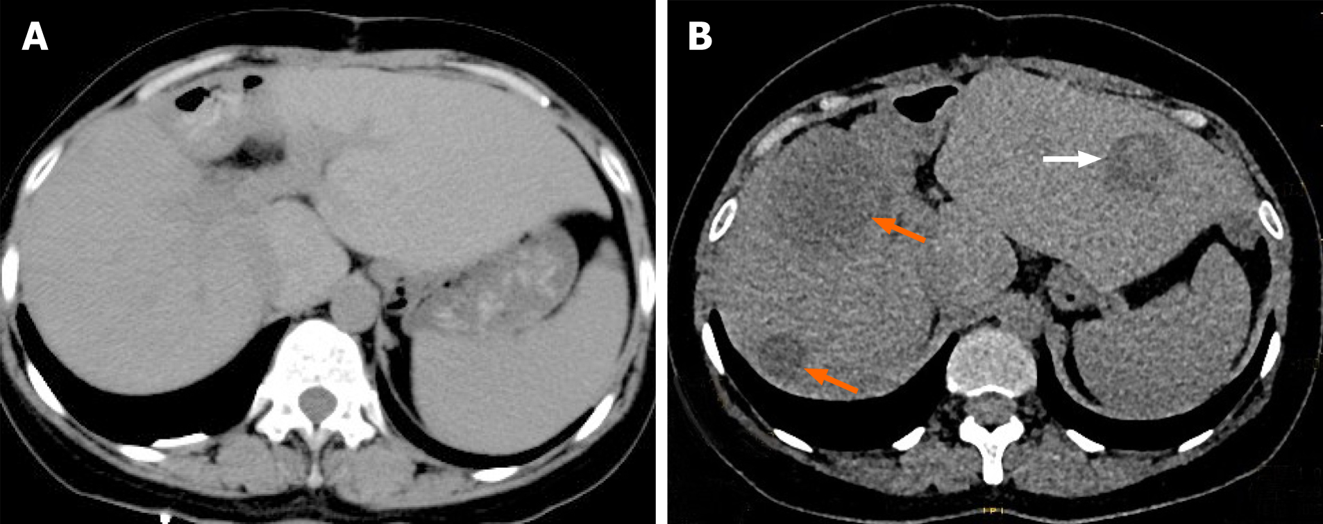
Figure 3 Postoperative re-examination images.
A: Computed tomography re-examination at 3 mo postoperatively did not indicate obvious abnormal density opacities in the liver parenchyma; B: Computed tomography re-examination at 1 year postoperatively revealed multiple patches of low- or slightly low-density opacities in the liver parenchyma (orange arrow), with patchy high-density hemorrhage in the lesion (white arrow).
- Citation: Ren SX, Li PP, Shi HP, Chen JH, Deng ZP, Zhang XE. Imaging presentation and postoperative recurrence of peliosis hepatis: A case report. World J Clin Cases 2021; 9(19): 5197-5202
- URL: https://www.wjgnet.com/2307-8960/full/v9/i19/5197.htm
- DOI: https://dx.doi.org/10.12998/wjcc.v9.i19.5197


