Copyright
©The Author(s) 2021.
World J Clin Cases. Jun 26, 2021; 9(18): 4810-4816
Published online Jun 26, 2021. doi: 10.12998/wjcc.v9.i18.4810
Published online Jun 26, 2021. doi: 10.12998/wjcc.v9.i18.4810
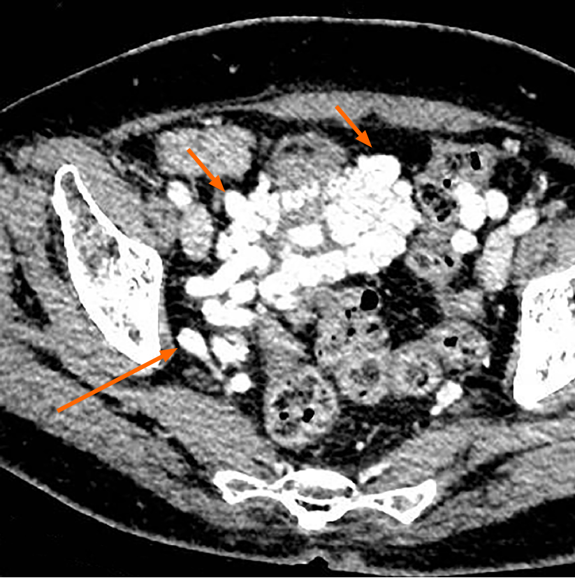
Figure 3 Axial contrast-enhanced computed tomography image demonstrating multiple tortuous and thickened veins on the anterior wall and both sidewalls of the bladder (short arrow).
The dilated vesical varices on the right side drained into the internal iliac vein (long arrow).
- Citation: Wei ZJ, Zhu X, Yu HT, Liang ZJ, Gou X, Chen Y. Severe hematuria due to vesical varices in a patient with portal hypertension: A case report. World J Clin Cases 2021; 9(18): 4810-4816
- URL: https://www.wjgnet.com/2307-8960/full/v9/i18/4810.htm
- DOI: https://dx.doi.org/10.12998/wjcc.v9.i18.4810


