Copyright
©The Author(s) 2021.
World J Clin Cases. Jun 26, 2021; 9(18): 4741-4747
Published online Jun 26, 2021. doi: 10.12998/wjcc.v9.i18.4741
Published online Jun 26, 2021. doi: 10.12998/wjcc.v9.i18.4741
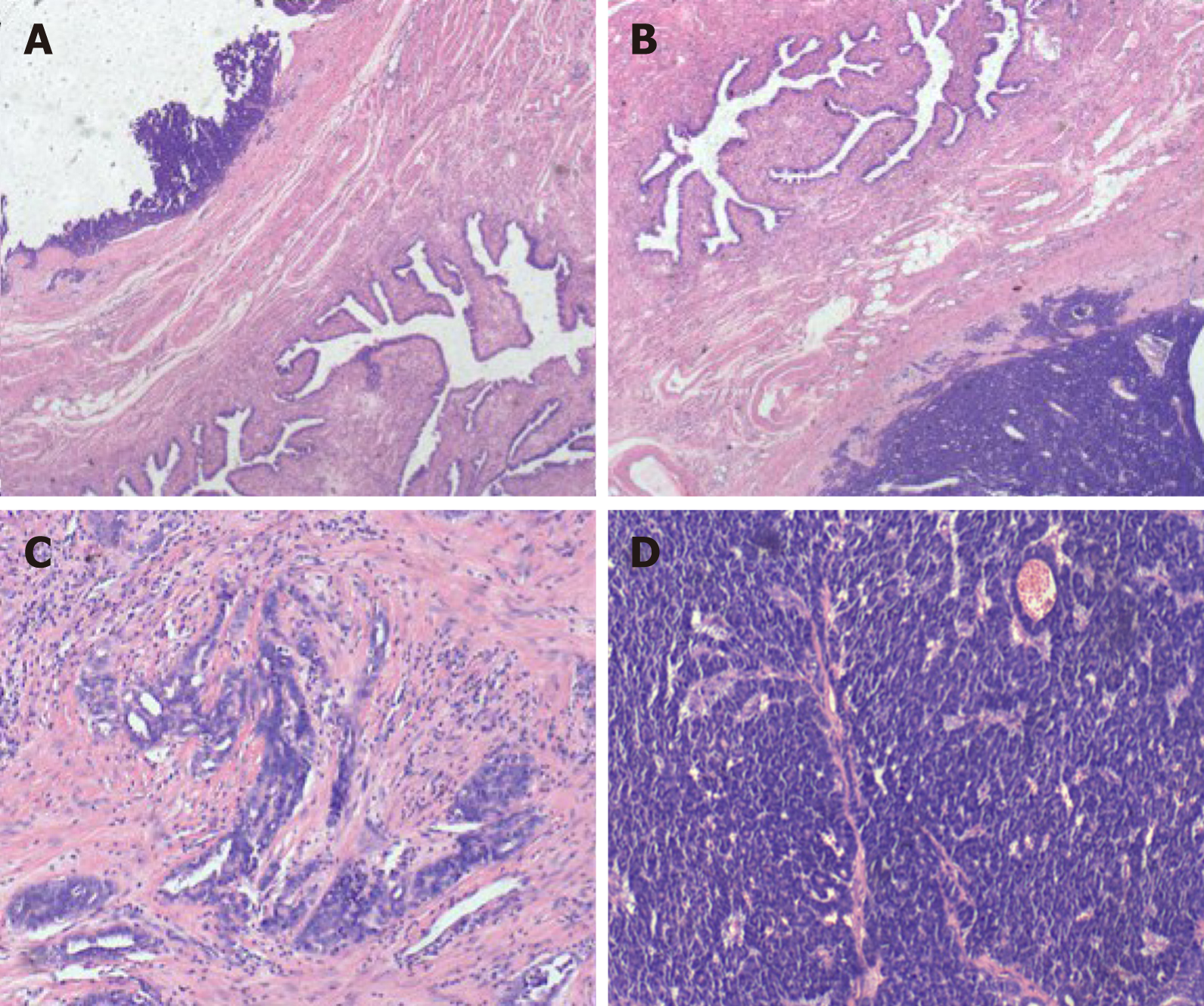
Figure 1 Histologic features of fallopian tube-mesonephric adenocarcinoma.
A and B: Pathological finding revealed that the tumor originated from the fallopian tube wall and involved the mucosa and serosal membrane of the fallopian tube; C and D: Vestiges of mesonephric hyperplasia (C) and hyperplasia into cancerous nests (D) were histologically found. The histologic patterns of the tumor were mainly solid.
- Citation: Xie C, Shen YM, Chen QH, Bian C. Primary mesonephric adenocarcinoma of the fallopian tube: A case report. World J Clin Cases 2021; 9(18): 4741-4747
- URL: https://www.wjgnet.com/2307-8960/full/v9/i18/4741.htm
- DOI: https://dx.doi.org/10.12998/wjcc.v9.i18.4741


