Copyright
©The Author(s) 2021.
World J Clin Cases. Jun 26, 2021; 9(18): 4734-4740
Published online Jun 26, 2021. doi: 10.12998/wjcc.v9.i18.4734
Published online Jun 26, 2021. doi: 10.12998/wjcc.v9.i18.4734
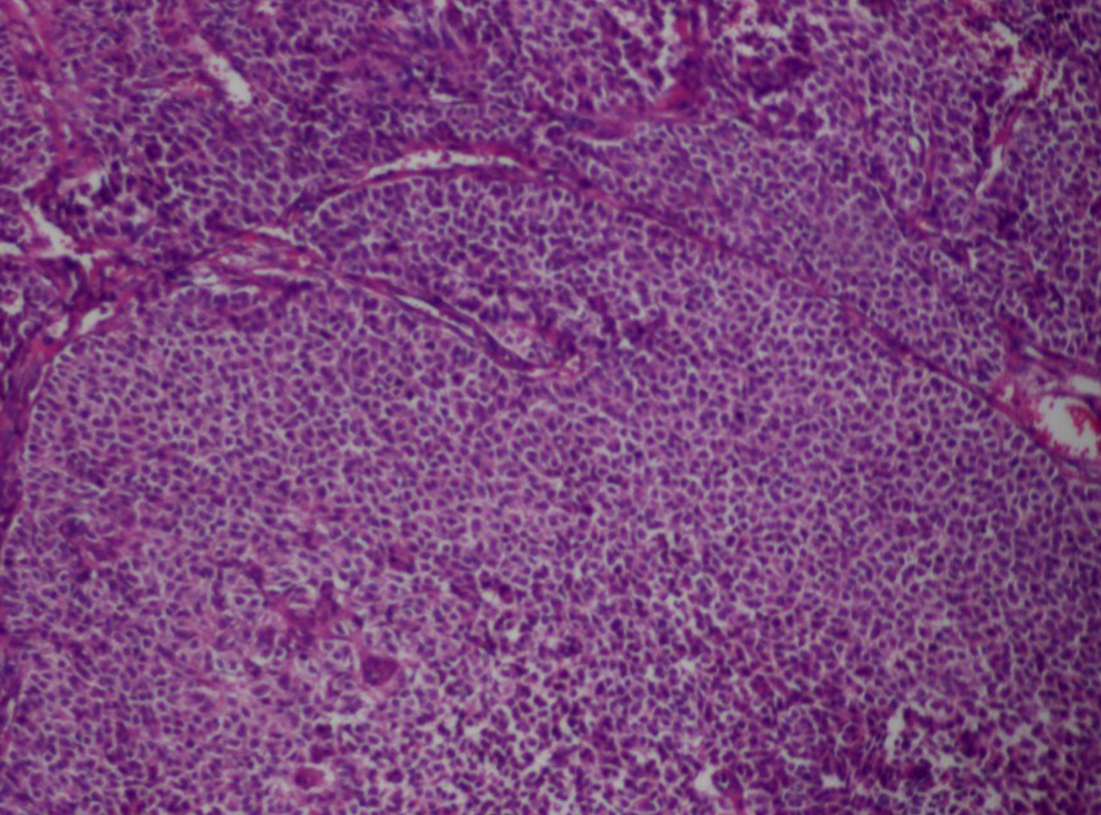
Figure 3 Pathology image showing round or oval cells of medium size, with a clear boundary, abundant cytoplasm, rounded nuclei, and visible karyokinesis (hematoxylin and eosin staining, × 200).
- Citation: Wu XJ, Xia HB, Jia BL, Yan GW, Luo W, Zhao Y, Luo XB. Meigs’ syndrome caused by granulosa cell tumor accompanied with intrathoracic lesions: A case report. World J Clin Cases 2021; 9(18): 4734-4740
- URL: https://www.wjgnet.com/2307-8960/full/v9/i18/4734.htm
- DOI: https://dx.doi.org/10.12998/wjcc.v9.i18.4734


