Copyright
©The Author(s) 2021.
World J Clin Cases. Jun 26, 2021; 9(18): 4734-4740
Published online Jun 26, 2021. doi: 10.12998/wjcc.v9.i18.4734
Published online Jun 26, 2021. doi: 10.12998/wjcc.v9.i18.4734
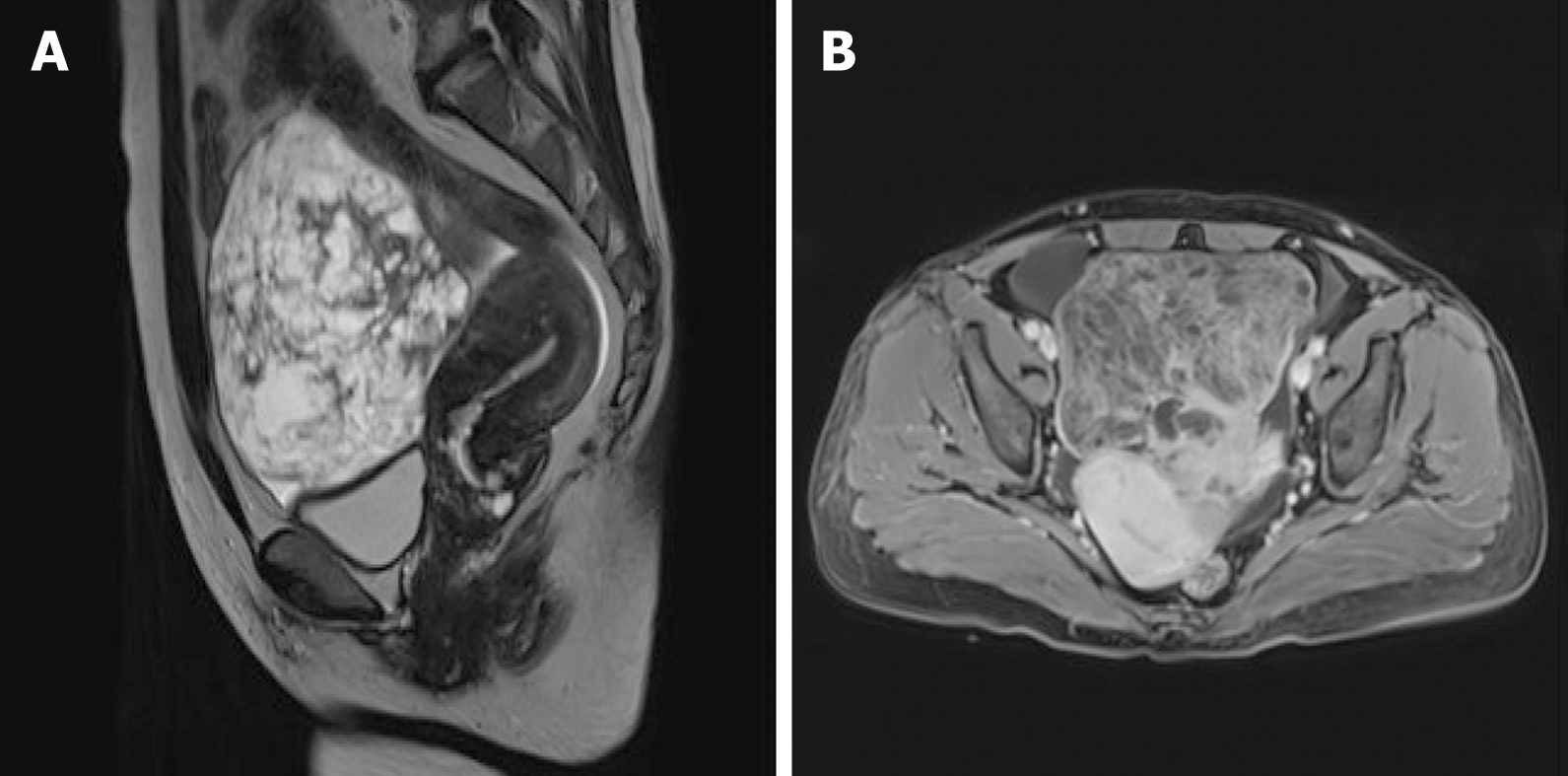
Figure 2 Pelvic magnetic resonance imaging.
A: T2-weighted pelvic magnetic resonance imaging (MRI) revealed a pelvic mass measuring about 11.6 cm × 10.0 cm × 12.4 cm with heterogeneous signal intensity and multiple hypointense separations with long T2 signals, a small amount of fat, characteristic signals of hemorrhage, and vessel signs; B: Contrast-enhanced MRI scan showed significant enhancement heterogeneity.
- Citation: Wu XJ, Xia HB, Jia BL, Yan GW, Luo W, Zhao Y, Luo XB. Meigs’ syndrome caused by granulosa cell tumor accompanied with intrathoracic lesions: A case report. World J Clin Cases 2021; 9(18): 4734-4740
- URL: https://www.wjgnet.com/2307-8960/full/v9/i18/4734.htm
- DOI: https://dx.doi.org/10.12998/wjcc.v9.i18.4734


